Louisiana State University School of Medicine New Orleans, LA
Farhan Mohiuddin, MD1, Ross Dies, MD2, Nali Gillespie, MD1, Rachel McMullen, DO1, Christine Bruins, MD1 1Louisiana State University School of Medicine, New Orleans, LA; 2Louisiana State University School of Medicine, Shreveport, LA Introduction: Epithelioid hemangioendothelioma (EHE) is a rare vascular tumor, accounting for less than 1% of all vascular neoplasms. Its estimated prevalence is 0.23–0.4 cases per million person-years. EHE most commonly affects the liver, lungs, and bones. The pathogenesis remains unclear, and it can be difficult to differentiate multicentric disease from primary lesions with metastasis without histopathologic and genetic evaluation. EHE is often an incidental finding, with many patients asymptomatic at diagnosis. Prognosis depends on disease location and features such as liver involvement and hilar metastases, with mean survival around 4.6 years. We present a case of EHE diagnosed after a patient presented with right upper quadrant (RUQ) pain.
Case Description/
Methods: A 24-year-old female with a history of diabetes, hyperlipidemia, and polycystic ovarian syndrome presented to the ED with five days of sharp RUQ pain radiating to the epigastrium. There were no clear exacerbating or relieving factors. She reported similar symptoms in 2023, but prior evaluation was reportedly unremarkable.
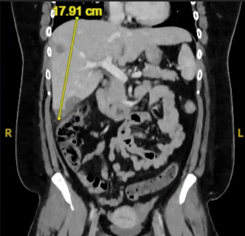
On presentation, her vital signs were stable. Labs revealed mild microcytosis and a slight elevation in alkaline phosphatase. CMP and CBC were otherwise unremarkable. CT of the abdomen and pelvis showed multiple bilateral pulmonary nodules, rim-enhancing hypoattenuating hepatic lesions, and reactive lymphadenopathy. Interventional radiology was consulted for a liver biopsy. Workup including tumor markers, infectious disease labs, and hematology-oncology evaluation was unrevealing. As the patient remained clinically stable, she was discharged pending biopsy results.
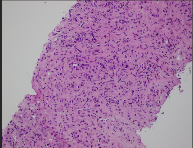
Pathology later revealed positive staining for CAMTA1, CD31, and ERG, and was negative for cytokeratin 7. Morphologic and immunohistochemical findings were diagnostic of epithelioid hemangioendothelioma. She was referred to hematology-oncology for further evaluation and treatment planning. Discussion: Due to its rarity, no standardized treatment protocol exists for EHE. Available options include surgical resection, chemotherapy, radiation, and in some cases, organ transplantation. EHE can mimic metastases, vascular malformations, granulomatous disease, and infections, complicating diagnosis. Its nonspecific presentation and lack of early biomarkers contribute to diagnostic delays. Greater clinical awareness and improved diagnostic tools are needed to aid earlier recognition and optimize outcomes in affected patients.
Figure: Figure 1: CT Abdomen Showing Liver Lesion
Figure: Figure 2: Histology of Biopsy Revealing Diagnosis
Disclosures: Farhan Mohiuddin indicated no relevant financial relationships. Ross Dies indicated no relevant financial relationships. Nali Gillespie indicated no relevant financial relationships. Rachel McMullen indicated no relevant financial relationships. Christine Bruins indicated no relevant financial relationships.
Farhan Mohiuddin, MD1, Ross Dies, MD2, Nali Gillespie, MD1, Rachel McMullen, DO1, Christine Bruins, MD1. P3878 - Epithelioid Hemangioendothelioma Presenting as Right Upper Quadrant Pain in a 24-Year-Old Female, ACG 2025 Annual Scientific Meeting Abstracts. Phoenix, AZ: American College of Gastroenterology.

.jpg "Farhan Mohiuddin, MD photo")