University of Mississippi Medical Center - - Jackson, MS Brandon, MS
Christopher G. Moody, DO1, Aneeq Muhammad Yousuf, MD2, Namrata Paladugu, MD2, Joydeep Chakraborty, MD2 1University of Mississippi Medical Center - - Jackson, MS, Brandon, MS; 2University of Mississippi Medical Center, Jackson, MS Introduction: Hereditary Hemorrhagic Telangiectasia (HHT) is an autosomal dominant disorder characterized by the formation of abnormal blood vessels in the skin, mucous membranes, brain, lungs, liver, and gastrointestinal tract. This condition results from ENG, ACVRL1, GDF4, or SMAD4 gene mutations and can lead to recurrent epistaxis, iron deficiency anemia, gastrointestinal bleeding, seizures, and pulmonary complications. Diagnosis requires a thorough history and physical exam and is made by meeting 3 of the 4 Curaçao Criteria, which include recurrent epistaxis, multiple telangiectasias, visceral lesions, and a positive family history.
Case Description/
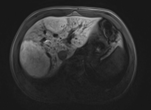
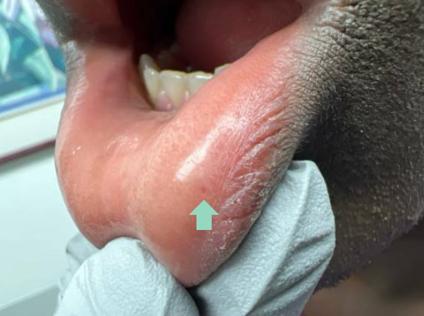
Methods: A 19-year-old African American male with a history of asthma, atopic dermatitis, Growth Hormone deficiency, and hypertension presented to Hepatology Clinic for evaluation of multiple atypical Focal Nodular Hyperplasia (FNH) lesions found on magnetic resonance imaging (MRI). At an Emergency Room visit for an episode of viral gastroenteritis, a computed tomography (CT) of his abdomen showed suspicion of liver lesions. An MRI obtained later showed multiple atypical FNH, with the largest measuring 8 centimeters in the right hepatic lobe (Figure 1). He was also found to have iron deficiency. He reported that he had repeated epistaxis since he was 13 to 14 years old, which previously required cauterization with Otolaryngology (ENT). Due to the known association of atypical FNH in HHT, a thorough physical exam was performed, revealing a single oral telangiectasia (Figure 2). Given his history and physical exam findings, he was referred to ENT, where he was found to have multiple nasal telangiectasias. CT head and MRI brain were obtained without evidence of AVMs. He met “Possible or Suspected HHT” per the Curaçao Criteria with recurrent epistaxis and multiple telangiectasias. Family history was unable to be obtained as he was adopted. He was referred to Hematology and Genetics for further workup, where his family wished to proceed with genetic testing to look for pathogenic sequence variants in ENG, ACVRL1, SMAD4, or GDF2 for further work-up. Discussion: In patients with atypical FNH lesions seen on imaging, it is important to have a high index of suspicion for HHT, especially if they report a history of recurrent epistaxis. A detailed history, physical examination, and utilizing the Curaçao Criteria are crucial in making the diagnosis if there is suspicion of HHT.
Figure: Multiple Atypical Focal Nodular Hyperplasia lesions found on MRI Abdomen.
Figure: Oral telangiectasia found during clinic visit with Hepatology.
Disclosures: Christopher Moody indicated no relevant financial relationships. Aneeq Muhammad Yousuf indicated no relevant financial relationships. Namrata Paladugu indicated no relevant financial relationships. Joydeep Chakraborty indicated no relevant financial relationships.
Christopher G. Moody, DO1, Aneeq Muhammad Yousuf, MD2, Namrata Paladugu, MD2, Joydeep Chakraborty, MD2. P3835 - Hereditary Hemorrhagic Telangiectasia: The Crucial Role of History and Physical Examination in Early Diagnosis, ACG 2025 Annual Scientific Meeting Abstracts. Phoenix, AZ: American College of Gastroenterology.