Brown University / Rhode Island Hospital Providence, RI
Alex Chang, MD, Sapana Gupta, MD, Kimberly Ho, MD, Nathan Sairam, MD, Amanda Pressman, MD, Fadlallah Habr, MD Brown University / Rhode Island Hospital, Providence, RI Introduction: Choledocholithiasis is uncommon after a cholecystectomy (CCY). The most prevalent cause is retained gallstones, presenting months after surgery. Initial presentation of choledocholithiasis multiple years after CCY is even rarer, with the longest-reported interval being 40 years. The current case series describes two cases of choledocholithiasis presenting over 10 years after CCY.
Case Description/
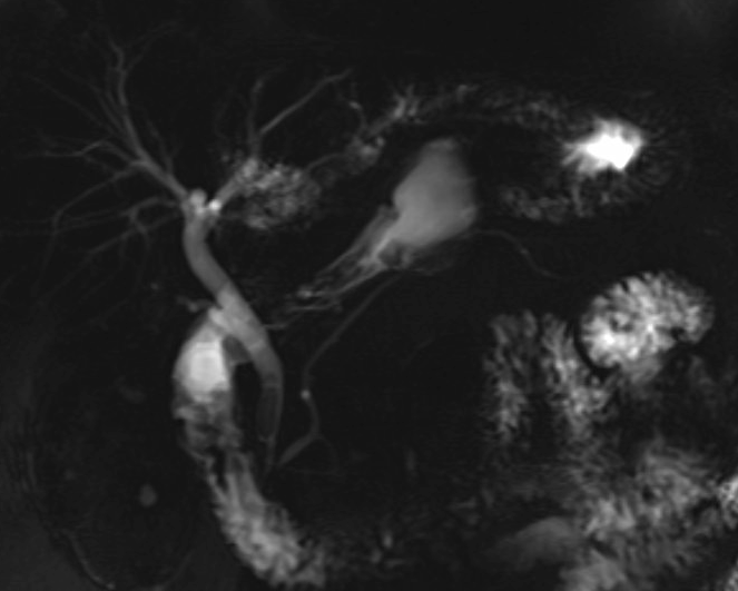
Methods: Case 1: A 76 year-old male patient presented to the ED with acute onset of abdominal pain in the right upper quadrant occurring after eating a meal. He reported that his pain came in waves lasting up to an hour. He had received a CCY 11-years prior. A CT-abdomen and pelvis demonstrated a surgically absent gallbladder. Subsequent MRCP demonstrated choledocholithiasis as well as intrahepatic ductal dilation (Figure 1).
ERCP was performed. The lower third of the CBD contained multiple stones. Complete removal of the stones was achieved with biliary sphincterotomy and balloon extraction. The patient reported resolution of symptoms with no post-procedural complications and was discharged successfully from the hospital after 2 days.
Case 2:
A 70 year-old female patient presented to the ED after waking up with left-sided chest pain radiating to her back. She denied an association of the pain with food intake. She had received a CCY in the 1970s, over 40 years ago. A CTA study was obtained to rule-out dissection but demonstrated a surgically absent gallbladder and biliary ductal dilation with gallstones.
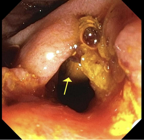
ERCP was performed. The CBD was markedly dilated with multiple gallstones, some > 1cm in size (Figure 2). Biliary sphincterotomy with balloon extraction was performed with removal of numerous stones, but multiple large stones remained. A plastic stent was placed into the CBD for biliary drainage. The patient was discharged from the hospital with plan for repeat ERCP with EHL which was ultimately successful. Discussion: To the best of our knowledge, the second case represents the longest described interval ( >40 years) between CCY and presentation of choledocholithiasis. Theories explaining late presentation of choledocholithiasis include migration of surgical clips, formation of stones in a remnant cystic duct, and possible primary stone formation in the CBD. The current cases reinforce that, while uncommon, choledocholithiasis can occur years after a CCY. Workup for choledocholithiasis should be pursued in patients with symptoms consistent with biliary colic even for patients with history of CCY.
Figure: Figure 1 MRCP demonstrating multiple small stones in the distal common bile duct
Figure: Figure 2 ERCP showing bile duct stone and sludge in the area of the papilla in the duodenum
Disclosures: Alex Chang indicated no relevant financial relationships. Sapana Gupta indicated no relevant financial relationships. Kimberly Ho indicated no relevant financial relationships. Nathan Sairam indicated no relevant financial relationships. Amanda Pressman indicated no relevant financial relationships. Fadlallah Habr indicated no relevant financial relationships.
Alex Chang, MD, Sapana Gupta, MD, Kimberly Ho, MD, Nathan Sairam, MD, Amanda Pressman, MD, Fadlallah Habr, MD. P4429 - Recurrent Gallstone Disease After Cholecystectomy: Two Cases of Choledocholithiasis in Patients More Than 10 Years After Cholecystectomy, ACG 2025 Annual Scientific Meeting Abstracts. Phoenix, AZ: American College of Gastroenterology.