Ivana Rubenstein, DO1, Mina Ayad, MD2, Aryama Sharma, MD3 1Broward Health Medical Center, Fort Lauderdale, FL; 2Broward Health North, Pompano Beach, FL; 3Broward Health North, Deerfield Beach, FL Introduction: Pancreatic cancer is a leading cause of cancer-related mortality, often diagnosed at advanced stages with limited therapeutic options. Tumor necrosis, infection, and abscess formation are common in these patients, particularly those undergoing chemotherapy. Pneumoperitoneum, the presence of air in the peritoneal cavity, is a rare but critical finding, frequently linked to gastrointestinal perforation.
Case Description/
Methods: A 77-year-old male with a history of pancreatic cancer undergoing chemotherapy, along with diabetes mellitus, hyperlipidemia, and obstructive sleep apnea, presented to the emergency department with worsening abdominal pain and fever over two days. On presentation, he was hypotensive, tachycardic, and tachypneic. Laboratory studies showed leukocytosis, elevated bilirubin, and transaminitis. CT abdomen and pelvis revealed a 3 cm necrotic pancreatic tumor with air in the superior left hepatic lobe, consistent with a ruptured tumor. Additionally, there was a 7 cm x 7 cm fluid collection, pneumoperitoneum, and air in the biliary tree. The patient was diagnosed with septic shock, secondary to peritonitis from the ruptured tumor. He was admitted to the ICU for intravenous fluids, pressor support, and broad-spectrum antibiotics. A CT-guided percutaneous drainage was performed, with cultures revealing Escherichia coli. After targeted antibiotic therapy, his clinical status improved. He was discharged with follow-up. Discussion: This case illustrates a rare but serious complication of pancreatic cancer: rupture of a necrotic tumor mass causing pneumoperitoneum and peritonitis. Advanced pancreatic tumors are prone to necrosis and infection due to their poor vascular supply. Tumor rupture contaminates the peritoneal cavity, leading to pneumoperitoneum and sepsis. The diagnosis was confirmed by CT imaging, which showed air in the peritoneal cavity and complex fluid collections. The presence of air in the biliary tree and pancreatic mass suggested biliary involvement, complicating management. Early recognition of pneumoperitoneum in patients with advanced pancreatic cancer is critical. Prompt imaging, appropriate antibiotic therapy, and interventional management are essential for improving outcomes, especially in immunocompromised patients undergoing chemotherapy.
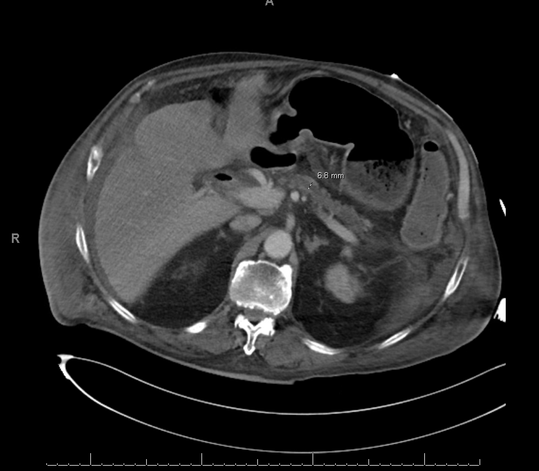
Figure: Figure 1: CT abdomen and pelvis revealing a 3 cm necrotic pancreatic tumor with air in the superior aspect of the left hepatic lobe, consistent with a ruptured tumor.
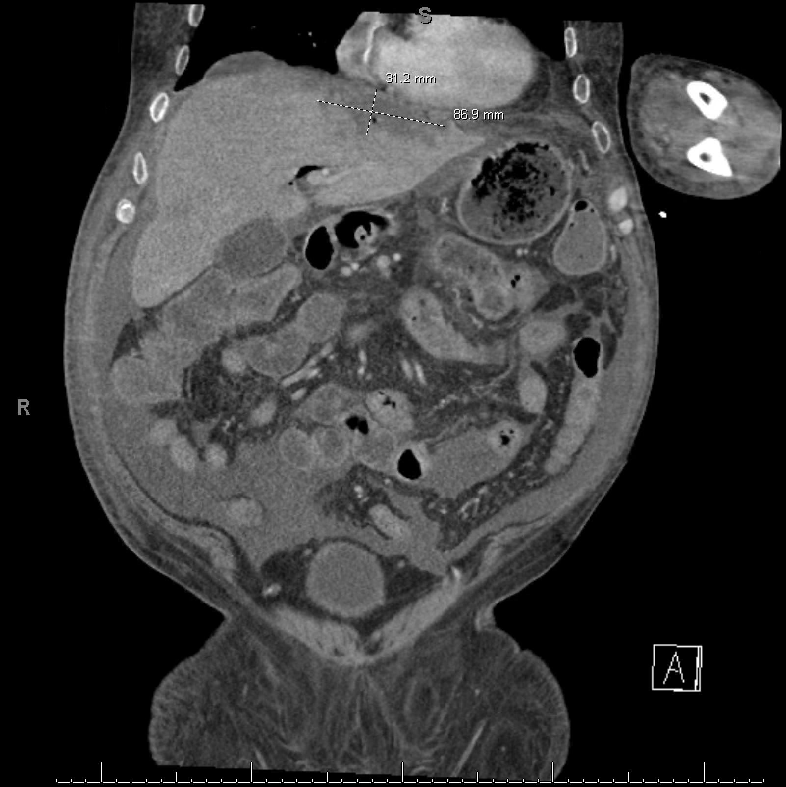
Figure: Figure 2: CT abdomen and pelvis demonstrating a 7 cm x 7 cm fluid collection, pneumoperitoneum, and air in the biliary tree.
Disclosures: Ivana Rubenstein indicated no relevant financial relationships. Mina Ayad indicated no relevant financial relationships. Aryama Sharma indicated no relevant financial relationships.
Ivana Rubenstein, DO1, Mina Ayad, MD2, Aryama Sharma, MD3. P4487 - Ruptured Pancreatic Tumor Complicated by Pneumoperitoneum and Sepsis in a Chemotherapy-Dependent Patient: A Case Report, ACG 2025 Annual Scientific Meeting Abstracts. Phoenix, AZ: American College of Gastroenterology.

 photo")