University of Central Florida, HCA Healthcare GME Gainesville, FL
Carson Creamer, DO1, Yeshika Thapa, MD2, Harsimran Kalsi, MD2, Kobina Essilfie-Quaye, MD2, Mohammad Abu Assi, MD2, Mohammed Janajri, MD2, Tony Brar, MD2, Yaseen Perbtani, DO2 1University of Central Florida, HCA Healthcare GME, Gainesville, FL; 2University of Central Florida, Gainesville, FL Introduction: Pancreatic ductal adenocarcinoma (PDAC) typically presents as a solid mass, often with associated ductal dilation and signs of local invasion. This case highlights a diagnostically challenging and atypical presentation of PDAC arising from intraductal papillary mucinous neoplasm (IPMN), mimicking multifocal pseudocysts in a patient with polysubstance use.
Case Description/
Methods: A 63-year-old male with a history of active polysubstance abuse presented with obstructive jaundice following a fall at home. Initial laboratory testing revealed elevated liver enzymes and total bilirubin. Magnetic resonance cholangiopancreatography demonstrated massive pancreatic and biliary dilation with a suspected multicystic mass in the pancreatic head. Endoscopic retrograde cholangiopancreatography (ERCP) showed severe common bile duct (CBD) stenosis, for which a 15 mm sphincterotomy and biliary sweep were performed. A plastic stent was placed, and brush cytology was negative for malignancy.
Further evaluation with endoscopic ultrasound (EUS) revealed multiple anechoic, septated, multicystic lesions throughout the pancreas, essentially replacing the entire gland, with no dominant solid mass. The endoscopic appearance was suggestive of multifocal pancreatic pseudocysts. Fine-needle aspiration of the cystic lesions demonstrated amylase < 20 U/L, glucose 51 mg/dL, carcinoembryonic antigen (CEA) of 5010 ng/mL, and cytology consistent with mucinous cystadenoma. CA 19-9 was markedly elevated at 1,810 U/mL.
Despite benign cytology and the absence of a discrete mass, the patient experienced progressive loss of pancreatic endocrine and exocrine function. Given the concern for malignant transformation, Surgical Oncology was consulted. The patient underwent total pancreatectomy with splenectomy, portal vein reconstruction, and cholecystectomy. Final pathology revealed moderately differentiated PDAC arising from IPMN with low- and high-grade dysplasia. The tumor involved the CBD, caused the biliary stricture, and demonstrated duodenal wall and perineural invasion. Discussion: This case is novel due to the unusual presentation of pancreatic adenocarcinoma as multifocal cystic lesions mimicking benign pseudocysts. It underscores the importance of thorough cyst fluid analysis and maintaining high clinical suspicion in patients with atypical cystic pancreatic disease and obstructive jaundice, even in the absence of overt malignancy on initial evaluation.
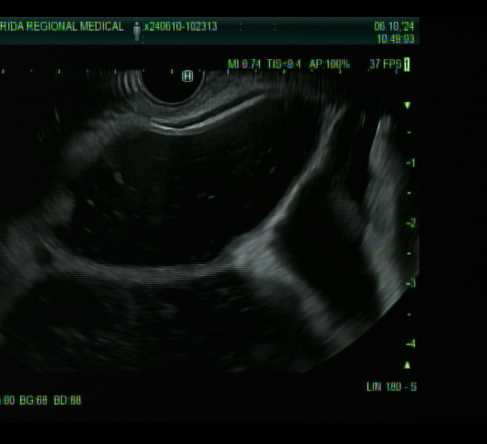
Figure: The EUS reveals multicystic and septated lesions throughout the pancreas, which were initially suggestive of multifocal pancreatic pseudocysts.
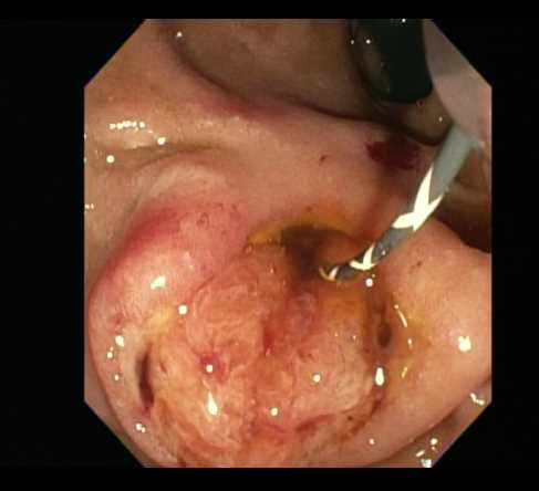
Figure: An endoscopic image of the ampulla of Vater during ERCP demonstrates abnormal morphology with mucosal irregularity and edema.
Disclosures: Carson Creamer indicated no relevant financial relationships. Yeshika Thapa indicated no relevant financial relationships. Harsimran Kalsi indicated no relevant financial relationships. Kobina Essilfie-Quaye indicated no relevant financial relationships. Mohammad Abu Assi indicated no relevant financial relationships. Mohammed Janajri indicated no relevant financial relationships. Tony Brar indicated no relevant financial relationships. Yaseen Perbtani indicated no relevant financial relationships.
Carson Creamer, DO1, Yeshika Thapa, MD2, Harsimran Kalsi, MD2, Kobina Essilfie-Quaye, MD2, Mohammad Abu Assi, MD2, Mohammed Janajri, MD2, Tony Brar, MD2, Yaseen Perbtani, DO2. P4482 - Atypical Presentation of Pancreatic Ductal Adenocarcinoma Mimicking Multifocal Pseudocysts, ACG 2025 Annual Scientific Meeting Abstracts. Phoenix, AZ: American College of Gastroenterology.