
Tuesday Poster Session
Category: Colon
 photo")
Andrew L. Petrou, MD (he/him/his)
Brown University / Rhode Island Hospital
Providence, RI
Melanosis coli (MC) is a benign pigmentation of the colonic mucosa, commonly associated with chronic use of anthraquinone laxatives like senna. The pigmentation results from lipofuscin accumulation in macrophages within the lamina propria, rather than melanin. Traditionally considered incidental, its impact on endoscopic visibility has recently come under scrutiny. Although some have suggested MC may obscure mucosal detail, others report improved polyp detection due to enhanced contrast between pigmented mucosa and non-pigmented lesions. We report a case where severe melanosis coli appeared to facilitate the identification of colonic polyps during colonoscopy, highlighting a potential diagnostic advantage of this often-overlooked condition.
Case Description/
Methods:
A 58-year-old patient with past medical history of chronic constipation and prior colonic polyps presented for screening colonoscopy. She was found to have melanosis coli throughout the entire length of the colon. Given the dark pigmentation the visibility of her polyps was more evident. She was found to have a hyperplastic polyp in the transverse colon as well as a cecal polyp and an ascending colon polyp that were found to be tubular adenomas with no evidence of dysplasia.
Discussion:
In our case, the severity of MC appeared to accentuate the visibility of polyps, suggesting a paradoxical diagnostic advantage. This observation aligns with prior studies demonstrating that MC may enhance colorectal adenoma detection by providing improved visual contrast between pigmented and non-pigmented mucosa. This contrast-enhancing effect resembles advanced imaging techniques such as narrow-band imaging or chromoendoscopy, which also rely on differential mucosal contrast to improve lesion identification. In this regard, extensive and uniform pigmentation seen in severe MC could function as a form of natural contrast aiding in the identification of subtle mucosal lesions. This case underscores the need to reassess assumptions about benign mucosal changes and consider their potential clinical value. While further research is warranted to quantify the influence of MC on adenoma detection rates, our findings contribute to a growing body of evidence suggesting that MC, under certain conditions, may assist rather than hinder colonoscopic evaluation.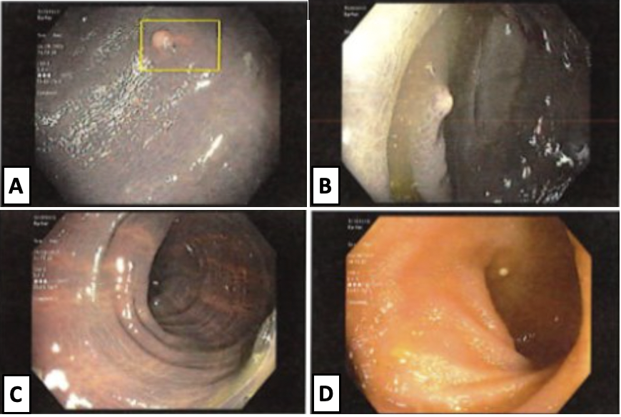
Figure: Figure 1. A. Ascending colonic polyp with background melanosis coli. B. Cecal polyp with background melanosis coli. C. Melanosis throughout the entire length of the colon. D. Normal appearing ileal mucosa.
Disclosures:
Andrew Petrou indicated no relevant financial relationships.
Grace Wilgucki indicated no relevant financial relationships.
Sean Fine indicated no relevant financial relationships.
Andrew L. Petrou, MD1, Grace Wilgucki, DO2, Sean Fine, MD1. P4606 - Pigment With a Perk: Severe Case of Melanosis Coli Unmasks Colonic Polyps, ACG 2025 Annual Scientific Meeting Abstracts. Phoenix, AZ: American College of Gastroenterology.