Mina Awadallah, MD, MSc1, Loc Ton, MD2, Viveksandeep Thoguluva Chandrasekar, MBBS3, Kenneth J.. Vega, MD, MHA4, John Erikson Yap, MD, MBA, FACG5 1University of Utah, Salt Lake City, UT; 2Kaiser Permanente, Sacramento, CA; 3Medical College of Georgia at Augusta University, Augusta, GA; 4Prisma Health Midlands, Columbia, SC; 5University of Utah Health, Salt Lake City, UT Introduction: Accidental foreign body ingestion is a common clinical occurrence, particularly in adults with dental prostheses, altered sensation, or distracted eating. Most ingested objects pass through the gastrointestinal (GI) tract without issue. However, sharp objects are associated with risk of mucosal injury or perforation. Toothpicks, though infrequently ingested and often not recalled by patients, pose a diagnostic challenge due to their radiolucent nature and potential to silently embed within the GI tract. We present a rare case of a toothpick discovered incidentally during a routine colonoscopy, with prior symptoms likely related to its lodgment.
Case Description/
Methods: A 53-year-old man with hypertension, type 2 diabetes, and obesity presented for his first screening colonoscopy. He reported no gastrointestinal symptoms and had no family history of colorectal cancer. Three weeks prior, he presented to the emergency department (ED) with acute abdominal pain and chills. Labs revealed leukocytosis (WBC 17,000/μL), but abdominal CT imaging was unremarkable. He was treated with intravenous fluids and Toradol, with resolution of symptoms. During colonoscopy, a wooden toothpick was discovered embedded in the ascending colon wall with surrounding purulent discharge. The foreign body was successfully retrieved using a snare without complication. A hemoclip was deployed to mark the site. Follow-up imaging showed no fluid collection or perforation, and the patient remained asymptomatic. On further questioning, he recalled potentially swallowing a toothpick while at church the day prior to his ED visit. Discussion: Sharp foreign bodies, such as toothpicks, can cause localized inflammation, perforation, or abscess formation if lodged within the gastrointestinal tract. The ascending colon is a rare site for such lodgment, and diagnosis is often delayed due to nonspecific symptoms and the lack of object radiolucency on standard imaging. In this case, the toothpick likely accounted for the patient’s prior abdominal symptoms, though it went undetected on CT. This highlights the diagnostic limitations in identifying ingested sharp objects and the importance of clinical suspicion in patients with unexplained abdominal symptoms. Endoscopic removal, when feasible, is an effective and safe therapeutic option.
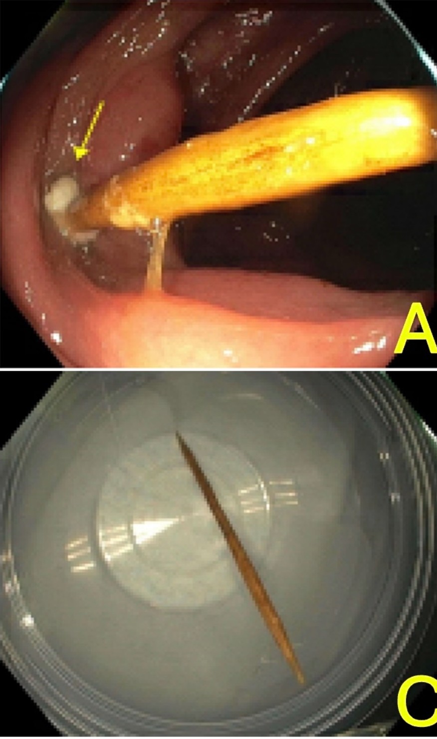
Figure: A.Embedded toothpick in the ascending colon with purulent discharge at the insertion site (yellow arrow). C.Retrieved toothpick following successful extraction.
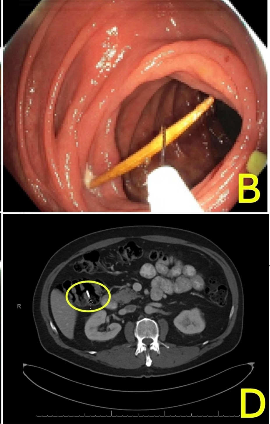
Figure: B.Endoscopic snare retrieval of the foreign body. D.Follow-up imaging demonstrating clip placement at the retrieval site, with no evidence of perforation or other complications(yellow circle).
Disclosures: Mina Awadallah indicated no relevant financial relationships. Loc Ton indicated no relevant financial relationships. Viveksandeep Thoguluva Chandrasekar indicated no relevant financial relationships. Kenneth Vega: Phathom Pharmceuticals – Speakers Bureau. John Erikson Yap: Phathom Pharmaceutical – Speakers Bureau. Steris – Consultant.
Mina Awadallah, MD, MSc1, Loc Ton, MD2, Viveksandeep Thoguluva Chandrasekar, MBBS3, Kenneth J.. Vega, MD, MHA4, John Erikson Yap, MD, MBA, FACG5. P4733 - Embedded and Undetected: A Rare Case of Toothpick Lodgment in the Ascending Colon, ACG 2025 Annual Scientific Meeting Abstracts. Phoenix, AZ: American College of Gastroenterology.