Mina Awadallah, MD, MSc1, Priscila Santiago, MD1, Gillian Hale, MD, MPH1, John Erikson Yap, MD, MBA, FACG2 1University of Utah, Salt Lake City, UT; 2University of Utah Health, Salt Lake City, UT Introduction: Colorectal cancer screening guidelines recommend surveillance colonoscopy for individuals with a personal history of polyps, primarily due to the elevated risk of malignancy associated with adenomatous polyps and sessile serrated lesions. However, there are no established follow-up protocols for other, less common polyp types. Mucosal leiomyomas are rare benign smooth muscle tumors of the gastrointestinal tract and are exceptionally uncommon in the colon. We present a rare case of a mucosal leiomyoma incidentally identified during routine surveillance colonoscopy in a high-risk patient.
Case Description/
Methods: A 78-year-old man with a medical history of hypertension,hyperlipidemia, and type 2 diabetes mellitus presented for high-risk colorectal cancer surveillance. He also had a significant personal history of colorectal polyps. Colonoscopy in 2017 revealed multiple polyps, including two cecal and two descending colon tubular adenomas, as well as two hyperplastic polyps in the sigmoid colon. A follow-up colonoscopy in 2020 identified one adenoma and one hyperplastic polyp in the sigmoid colon; all lesions were completely removed with no evidence of dysplasia or malignancy.
Despite his advance age, the patient remains highly functional and independent. He has previously discussed stopping surveillance; however, given his strong family history of longevity—with several relatives living beyond 100 years—he opted to proceed with one final surveillance colonoscopy in 2025. During this procedure, a 5mm sigmoid polyp was resected with a cold snare, and histopathology revealed a mucosal leiomyoma, a rare benign spindle cell tumor arising from the muscularis mucosae. Immunohistochemical staining was performed to exclude gastrointestinal stromal tumor (GIST). The spindle cells stained diffusely positive for desmin, consistent with smooth muscle origin, while CD117 and DOG1 were negative, effectively ruling out GIST.
Discussion: This case highlights the importance of recognizing rare polyp types beyond adenomatous, sessile serrated, and hyperplastic lesions. A mucosal leiomyoma,an uncommon histologic finding,was incidentally discovered during routine colorectal cancer surveillance. While most colonic leiomyomas are submucosal and rectosigmoid in location, mucosal-based tumors are rare. Histologic evaluation with immunohistochemistry is essential to distinguish benign mesenchymal lesions from entities like GIST, ensuring appropriate management and avoiding overtreatment.
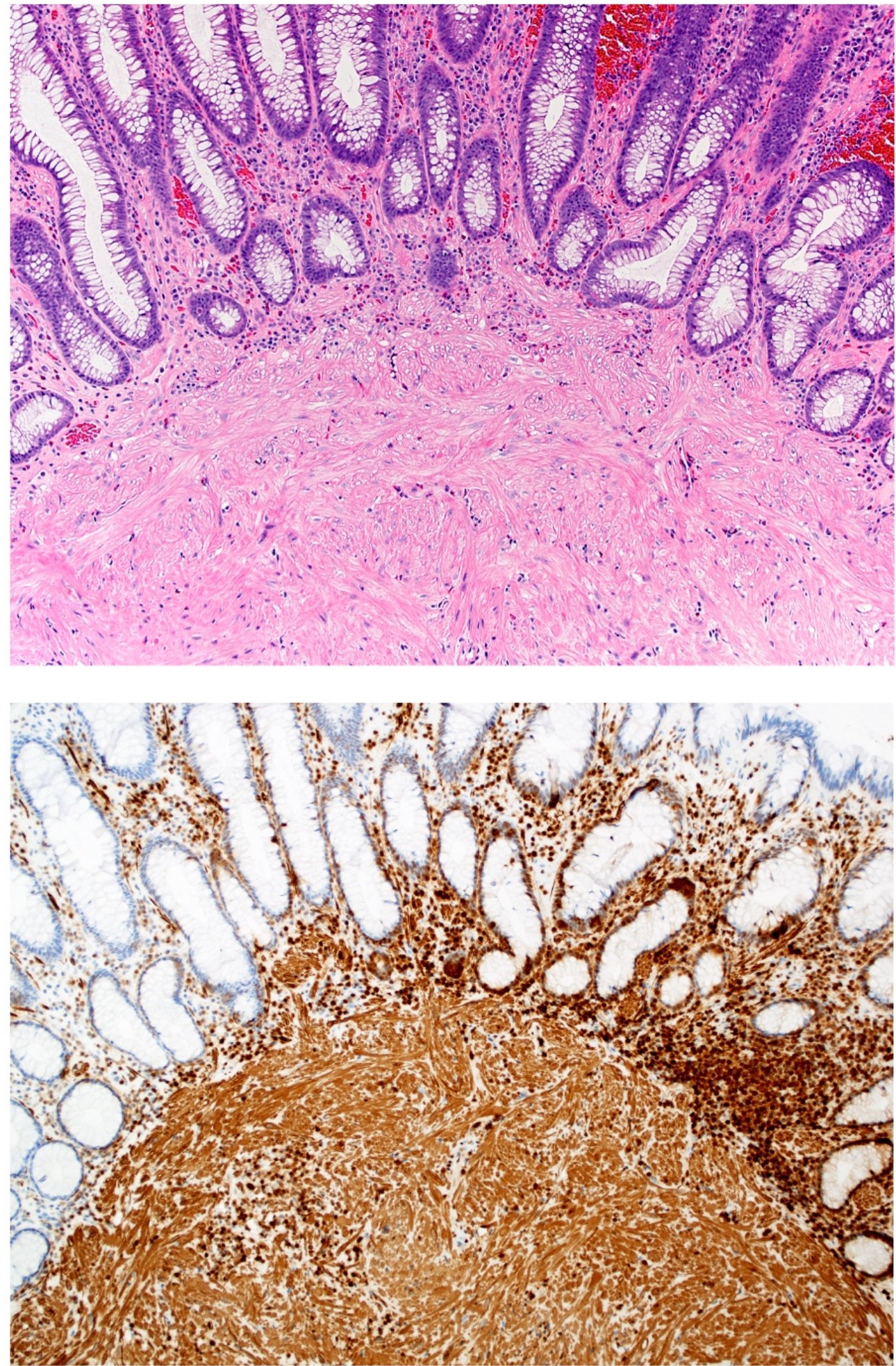
Figure: Sections of the polyp show a disorganized proliferation of bland spindled cells with brightly eosinophilic cytoplasm arising from the muscularis mucosae (H+E stain, 10X). Desmin immunostain for smooth muscle shows diffuse strong expression throughout the lesion. Stains for CD117 and DOG1 were negative, excluding a gastrointestinal stromal tumor (10X magnification).
Figure: Endoscopic image of the sigmoid colon (yellow arrows pointing to the polyp)
Disclosures: Mina Awadallah indicated no relevant financial relationships. Priscila Santiago indicated no relevant financial relationships. Gillian Hale indicated no relevant financial relationships. John Erikson Yap: Phathom Pharmaceutical – Speakers Bureau. Steris – Consultant.
Mina Awadallah, MD, MSc1, Priscila Santiago, MD1, Gillian Hale, MD, MPH1, John Erikson Yap, MD, MBA, FACG2. P4732 - Beyond Adenomas: An Uncommon Mucosal Leiomyoma in the Sigmoid Colon, ACG 2025 Annual Scientific Meeting Abstracts. Phoenix, AZ: American College of Gastroenterology.