
Tuesday Poster Session
Category: Colon
Marisa Pope, DO
Jefferson Health
Cherry Hill, NJ
Endometriosis is the existence of endometrial tissue implants outside of the uterus that can occur on other solid organs within the body. Endometriosis most commonly occurs within the rectosigmoid junction within the bowel and only occurs 2-5% of the time within the cecum. The symptoms of endometrial implants within the bowel can present as chronic abdominal pain, hematochezia, constipation, diarrhea, dyspareunia. We present a case of cecal endometriosis masquerading as a colonic tumor in appearance.
Case Description/
Methods:
A 46 year old female with a past medical of endometriosis s/p hysterectomy, chronic constipation who initially presented to the hospital for evaluation of abdominal in the LLQ. During her hospitalization she had a CT abdomen/pelvis with contrast that showed a cecal mass. Subsequently, she underwent a colonoscopy that showed a thickened fold in the cecum that was biopsied revealing colonic mucosa with mild chronic active colitis. Patient was discharged to follow up with gastroenterology and hepatology for MRI abdomen with and without contrast to further evaluation of her cecal mass. She underwent an MRI abdomen with and without contrast that revealed a persistent mass like abnormality in the right lower quadrant and hepatic adenoma. She then underwent a PET scan to further evaluate the lesion that noted a hypermetabolic mass in the right lower quadrant that was suspicious of being a small bowel neuroendocrine tumor or GIST. Patient then followed up with general surgery for an exploratory laparotomy with ileal resection, partial colectomy with primary anastomosis for mass of the cecum. After the mass was resected they found it to be consistent with endometriosis and mass in the segment VII of the liver.
Discussion: Our patient has a known history of endometriosis that had previously been diagnosed and treated with a laparotomy and hysterectomy. Our case is unique as the patient had recurrent endometriosis presenting in the cecum. Initially it was a diagnostic challenge that was unable to be diagnosed without surgery and was concerned to be a GIST or neuroendocrine tumor. Our patient had non specific bite on bite biopsies showing inflammation but did not reveal the diagnosis. Despite patient's subsequent imaging the final diagnosis was ultimately made due to histopathologic analysis after surgical resection. It is important to recognize the diagnostic challenge of recognizing cecal or bowel endometriosis given it’s low occurrence.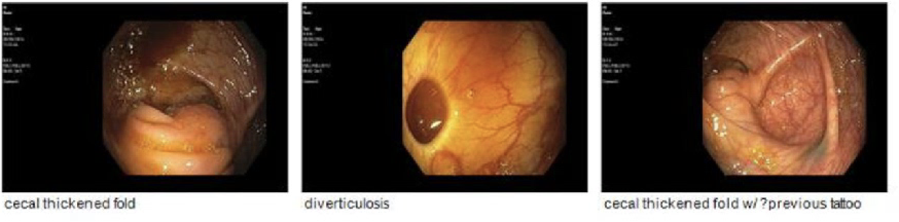
Figure: Cecal fold thickening
Disclosures:
Marisa Pope indicated no relevant financial relationships.
Joann Ha indicated no relevant financial relationships.
Mansi Sheth indicated no relevant financial relationships.
Seth Lipshutz indicated no relevant financial relationships.
Drew Chiesa indicated no relevant financial relationships.
Marisa Pope, DO1, Joann Ha, DO2, Mansi Sheth, DO3, Seth Lipshutz, DO4, Drew Chiesa, DO5. P4729 - Case of Cecal Endometriosis a Diagnostic Challenge That Requires a Multidisciplinary Approach, ACG 2025 Annual Scientific Meeting Abstracts. Phoenix, AZ: American College of Gastroenterology.