Paola Pena, MD, Qusai AlMasad, MD, Aryanna Sousa, MD, MPH, Thaer Abdelfattah, MD, MPH Roger Williams Medical Center, Providence, RI Introduction: Surgical clip migration into the common bile duct (CBD) is a rare but delayed complication of laparoscopic cholecystectomy. Clips serve as ideal environments for stone formation, imitating choledocholithiasis, which can happen early or even decades after surgery. We present a case of surgical clip migration causing biliary obstruction more than 20 years after surgery.
Case Description/
Methods: A 64-year-old female with a medical history of hypertension and surgical history of cholecystectomy 23 years prior who presented to the hospital with two days of worsening right upper quadrant, epigastric abdominal pain radiating to the back, associated with nausea, vomiting, and decreased oral intake.
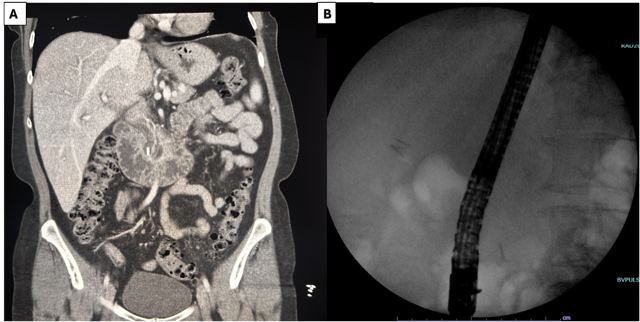
On admission, labs revealed elevated liver function tests [T. bilirubin: 3.6mg/dL (0.2-1.0mg/dL), AST:209u/L (10-42u/L), ALT:329u/L (10-60u/L), Alkaline phosphatase 170u/L (42-121u/L)] , blood cultures grew E. Coli and Klebsiella Oxytoca. Abdominal CT demonstrated intrahepatic and extrahepatic biliary duct dilation, with the CBD measuring up to 10 mm. A linear, 9 mm high-density object was identified in the distal CBD, which was presumed to be a migrated surgical clip.
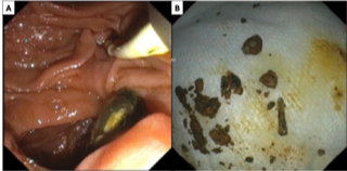
The patient underwent endoscopic retrograde cholangiopancreatography (ERCP), which confirmed choledocholithiasis. Biliary sphincterotomy and balloon extraction were performed, resulting in complete stone removal. The stone retrieved corresponded to the radiopaque clip seen on fluoroscopy; the stone was manually broken down, revealing a surgical clip inside. Post-procedure, the patient's symptoms resolved, and liver chemistries normalized. Discussion: In this case, the diagnosis was suspected by imaging and confirmed with ERCP. The case emphasized the importance of considering late surgical complications such as post-cholecystectomy clip migration in the differential diagnosis of obstructive biliary pathologies or even cholangitis, which can happen even decades after the initial procedure was performed. Timely recognition and endoscopic management are critical for resolution, which usually recovers without chronic complications.
Figure: Figure 1A: Visualization of surgical clip in the CBD. Figure 1B: Visualization of clip during ERCP before contrast injection.
Figure: Figure 2A: Stone visualization and removal. Figure 2B: Broken down stone revealing surgical clip.
Disclosures: Paola Pena indicated no relevant financial relationships. Qusai AlMasad indicated no relevant financial relationships. Aryanna Sousa indicated no relevant financial relationships. Thaer Abdelfattah indicated no relevant financial relationships.
Paola Pena, MD, Qusai AlMasad, MD, Aryanna Sousa, MD, MPH, Thaer Abdelfattah, MD, MPH. P5727 - Clip-Colelithiasis: A Case of Surgical Clip Migration Presenting as Choledocholithiasis Two Decades Post-Cholecystectomy, ACG 2025 Annual Scientific Meeting Abstracts. Phoenix, AZ: American College of Gastroenterology.