Julie Lee, DO1, Faisal Mehmood, MD1, Bassel Al-Lahham, MD, FACG1, Gavin Levinthal, MD2 1HonorHealth, Scottsdale, AZ; 2Honor Health Gastroenterology - SCOTTSDALE, AZ, Scottsdale, AZ Introduction: Gastrointestinal (GI) xanthomas are rare, benign lesions characterized histologically by lipid-laden foamy histiocytes within the lamina propria. Gastric xanthomas are typically discovered incidentally during endoscopy, with reported prevalence rates ranging from 0.2% to 7% in various studies. While gastric xanthomas have been associated with premalignant conditions and even rapid progression of gastric cancer, the clinical relevance of colorectal xanthomas remains uncertain. We present a patient who was seen for a routine screening colonoscopy that led to a diagnosis of multifocal gastrointestinal xanthomatosis.
Case Description/
Methods: A 50-year-old woman with a past medical history of iron deficiency anemia and GERD presented for a routine colon cancer screening. Colonoscopy revealed multiple white plaques throughout the colon. Biopsies of those plaques confirmed mucosal xanthomas. Later that evening, she presented with melena, lightheadedness, and left-sided abdominal pain. She denied NSAID or anticoagulant use. Her hemoglobin was 10.2 g/dL (previously 13.1 g/dL a year prior). The lipid panel did not show elevated LDL levels. Upper endoscopy revealed white plaques diffusely in the stomach. Gastric biopsy confirmed mucosal xanthomas; H. pylori immunohistochemistry was negative. She had a similar GI bleed in the past, and the workup at that time was unremarkable. Discussion: The etiology of GI xanthomas is not fully understood. Chronic mucosal inflammation, oxidative stress, H. pylori infection, metabolic disorders such as diabetes mellitus, and dyslipidemia have all been postulated as contributing factors. Although most xanthomas are considered benign, gastric xanthomas have been independently associated with rapid gastric cancer growth, suggesting they may reflect a field effect of mucosal vulnerability. The bleeding risk associated with GI xanthomas is considered low but has been reported, particularly in the setting of mucosal friability or ulceration due to vascular defects – this may explain the melena our patient experienced post-colonoscopy. Diffuse GI xanthomatosis is a rare and underrecognized condition that may signal chronic mucosal injury. While colorectal xanthomas remain of uncertain significance, their presence in patients with unexplained GI symptoms or bleeding should prompt further investigation. Long-term studies are needed to better characterize their prognostic implications and inform surveillance strategies.
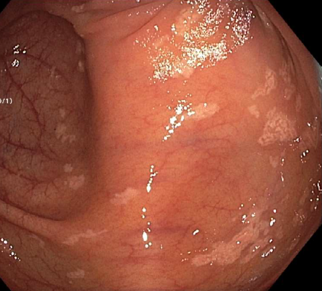
Figure: Image from the colonoscopy demonstrating multiple small areas of white discoloration/white plaques scattered in the cecum.
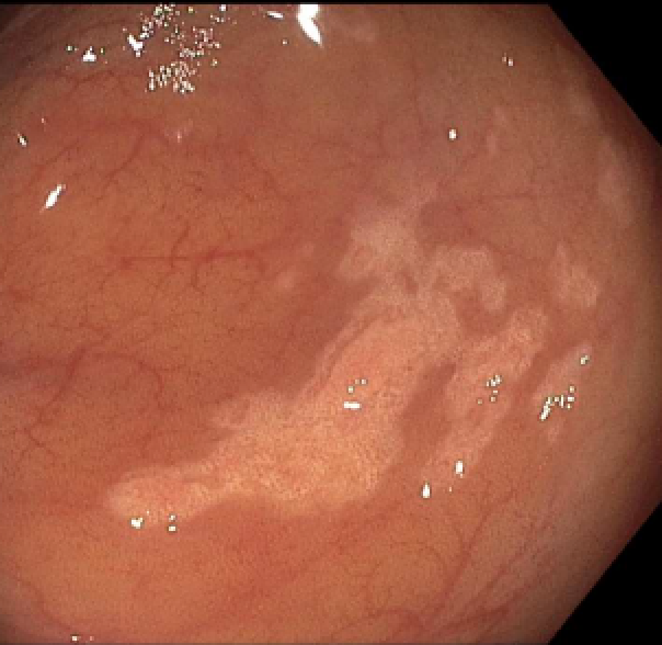
Figure: Additional area of white discoloration/white plaque in the cecum.
Disclosures: Julie Lee indicated no relevant financial relationships. Faisal Mehmood indicated no relevant financial relationships. Bassel Al-Lahham indicated no relevant financial relationships. Gavin Levinthal indicated no relevant financial relationships.
Julie Lee, DO1, Faisal Mehmood, MD1, Bassel Al-Lahham, MD, FACG1, Gavin Levinthal, MD2. P5280 - Diffuse Gastrointestinal Xanthomatosis Revealed During Routine Cancer Screening, ACG 2025 Annual Scientific Meeting Abstracts. Phoenix, AZ: American College of Gastroenterology.