Garrett Teskey, DO1, Oneka Daniels, MD2, Zoilo Placeres-Leon, MD2, Mazer Ally, MD3 1Scripps Mercy Hospital, Yorba Linda, CA; 2Georgetown Public Hospital Corporation, Georgetown, Demerara-Mahaica, Guyana; 3Scripps Clinic Medical Group, San Diego, CA Introduction: Foreign body (FB) ingestion is usually benign, but sharp objects can cause life-threatening complications if they perforate the esophagus. Cervical esophageal perforation, though rare, may result in abscess, mediastinitis, or fistula formation. Prompt recognition and intervention are essential.
We present a rare case of proximal esophageal perforation caused by a chicken bone with delayed diagnosis, leading to a cervical abscess and esophagocutaneous fistula requiring surgical repair.
Case Description/
Methods: A 45-year-old woman with no significant history presented repeatedly over several days with sore throat, dysphagia, low-grade fevers, weight loss, and a palpable neck mass. She reported swallowing a chicken bone, but initial evaluations were unremarkable, and she was repeatedly discharged with conservative treatment.
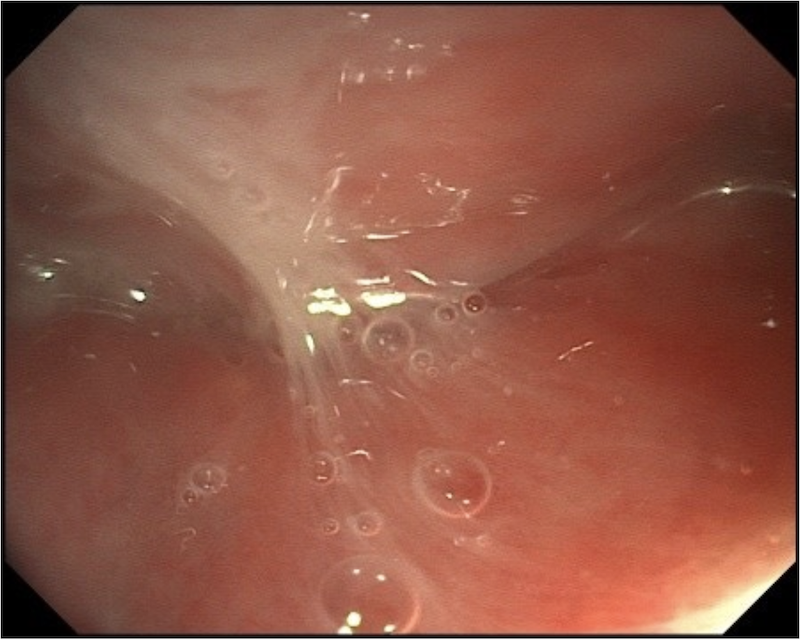
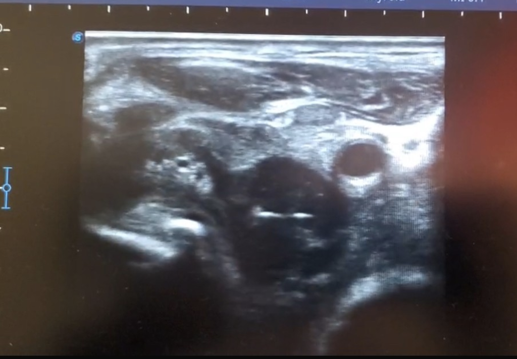
Persistent symptoms prompted gastroenterology referral. Exam revealed left cervical swelling and tenderness. Upper endoscopy displayed purulent drainage in the esophagus accompanied by a foul odor. Biopsies showed chronic gastritis, negative for H. pylori. Point-of-care ultrasound (POCUS) revealed a complex fluid collection with air and a suspected foreign body outside the esophageal wall. Aspiration yielded purulent fluid, raising concern for esophagocutaneous fistula.
Surgical exploration confirmed a deep neck abscess and retrieved a chicken bone fragment. The esophagus was repaired using a muscle flap, and a gastrostomy tube was placed for enteral feeding. Gastric decompression was initiated with a nasogastric tube. The patient recovered well, tolerated gastrostomy feeds, and a barium swallow showed no leak. She was discharged in stable condition with plans for gastrostomy removal after one month. Discussion: This case highlights the diagnostic challenge of esophageal perforation, especially with subtle or delayed symptoms. Cervical perforations are particularly dangerous due to proximity to vital structures. POCUS was pivotal in identifying the abscess and suggesting a retained FB, highlighting its utility in early evaluation when other imaging is inconclusive.
Despite delayed intervention, appropriate clinical suspicion and multidisciplinary mediation can achieve excellent outcomes, as demonstrated by this patient’s recovery and successful discharge.
Figure: Point-of-care ultrasound (POCUS) revealing a complex fluid collection with associated foreign body outside the esophageal wall.
Figure: Upper endoscopy showing purulent drainage in the esophagus.
Garrett Teskey, DO1, Oneka Daniels, MD2, Zoilo Placeres-Leon, MD2, Mazer Ally, MD3. P5010 - Chicken Bone Saga: Esophageal Perforation by Chicken Bone Causing Cervical Abscess and Esophagocutaneous Fistula: A Case Report, ACG 2025 Annual Scientific Meeting Abstracts. Phoenix, AZ: American College of Gastroenterology.