Ese Uwagbale, MD1, Venkat Arutla, MD2, Kevin Casey, MD2 1Rochester General Hospital, Webster, NY; 2Rochester General Hospital, Rochester, NY Introduction: Adenosquamous carcinoma (ASC) of the esophagus is a rare type of esophageal cancer that contains both adenocarcinoma and squamous cell carcinoma elements. ASC of the esophagus has a poorer prognosis and a higher rate of lymph node and distant metastasis compared to adenocarcinoma or squamous cell carcinoma alone. We present a case of ASC of the esophagus in a 61-year-old man.
Case Description/
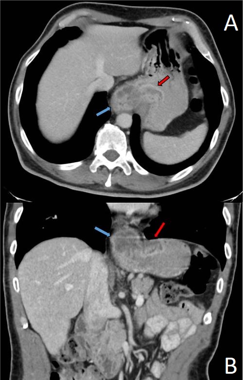
Methods: A 61-year-old male with a past medical history of intellectual disability and chronic cigarette smoker presented to the ED with 2-month history of vomiting after meals and 27 pounds weight loss. CT Abdomen and Pelvis revealed a 1.9cm thickening of the gastroesophageal (GE) junction (Figure 1), suggesting possible malignancy. Upper endoscopy showed near complete obstruction at the lower esophagus at 39 cm with friable mucosa concerning malignancy, which was biopsied. Pathology showed moderately differentiated invasive carcinoma with both gland formation and non-keratinizing squamous differentiation (Figure 2), which is most consistent with adenosquamous carcinoma. A gastrojejunostomy tube was placed for nutrition. The patient was initially treated with Carboplatin and Paclitaxel-based neoadjuvant chemoradiation with curative intent. However, he developed metastatic disease and declined further treatment with palliative chemotherapy and immunotherapy. The patient's family decided for him to be made comfort care. Discussion: ASC of the esophagus is a rare type of esophageal cancer, accounting for about 1% of all esophageal carcinomas. A multidisciplinary approach is required for treatment. Its prognosis is difficult to ascertain given its low incidence, which is reflected in current literature with conflicting data. ASC has the potential to have worse prognosis, with some studies suggesting lower mean survival time compared to esophageal squamous cell carcinoma or esophageal adenocarcinoma alone. Prompt diagnosis of this cancer is essential in guiding neoadjuvant chemotherapy, radiation, surgical resection, and potential immunotherapy to augment patients' prognosis.
Pathology slides courtesy of Dr. Joseph Hatem (pathology department Rochester General Hospital).
Figure: Figure 1: Gastroesophageal junction thickening (blue arrow) with extension into gastric cardia (red arrow) on axial (A) and sagittal (B) planes.
Figure: Figure 2: Hematoxylin and Eosin stain of ASC showing squamous component (A), with positive p40 immunohistochemical stain in squamous component (B), and adenocarcinoma component (C).
Disclosures: Ese Uwagbale indicated no relevant financial relationships. Venkat Arutla indicated no relevant financial relationships. Kevin Casey indicated no relevant financial relationships.
Ese Uwagbale, MD1, Venkat Arutla, MD2, Kevin Casey, MD2. P4974 - Double Trouble: A Rare Esophageal Cancer, ACG 2025 Annual Scientific Meeting Abstracts. Phoenix, AZ: American College of Gastroenterology.

 photo")