Samuel Schachter, MD, Alexandra Selby, MD, Mark Malamood, MD, Nicholas McDonald, MD Temple University, Philadelphia, PA Introduction: Esophageal epidermoid metaplasia (EEM) is a rare and underrecognized condition with malignant potential. Here, we present a case of esophageal dysphagia caused by stricture from circumferential EEM.
Case Description/
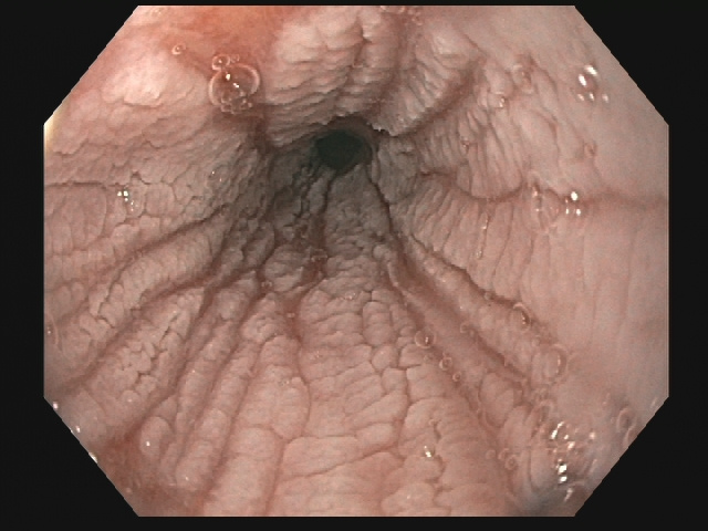
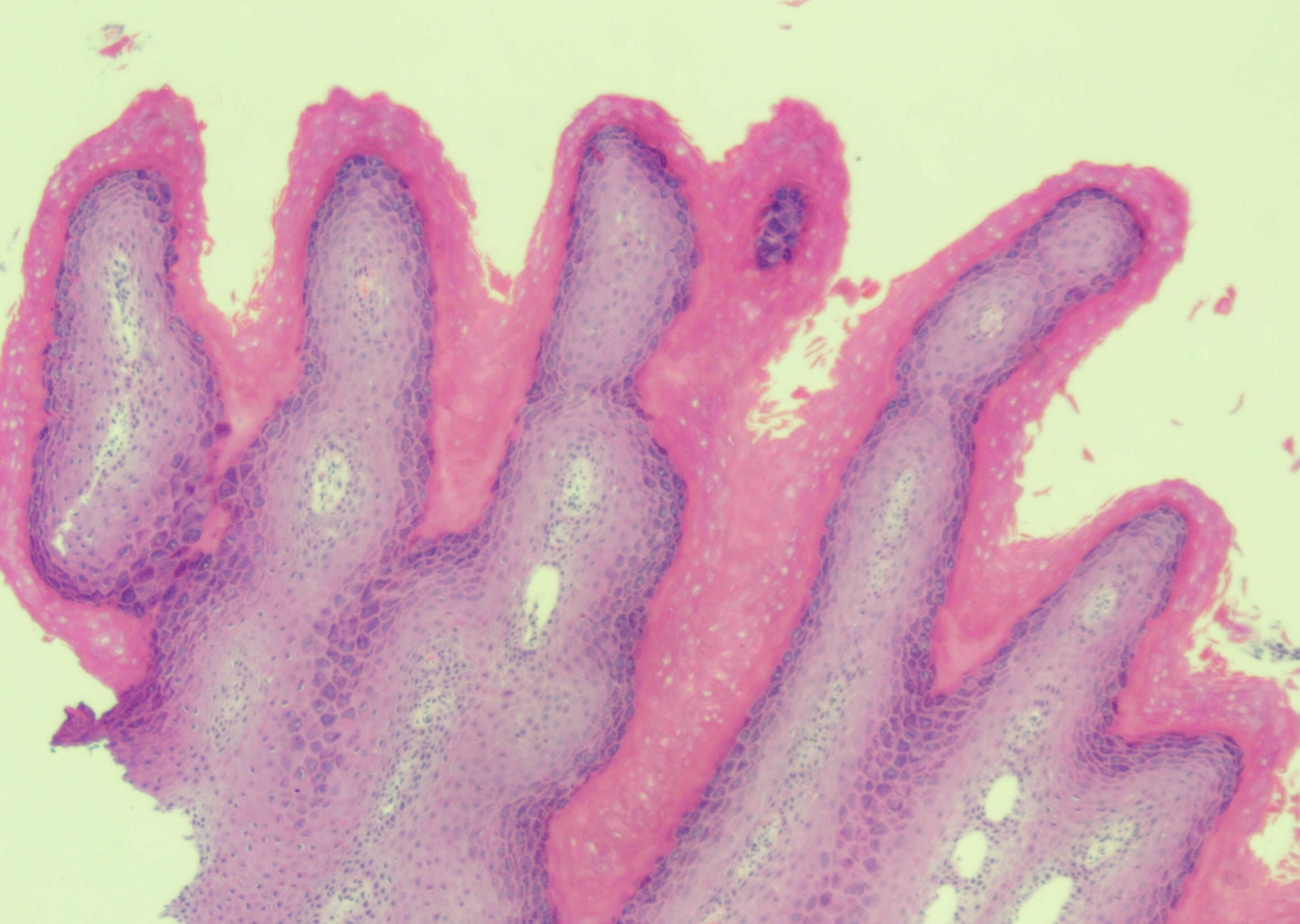
Methods: A 63-year-old man presented with acute on chronic progressive dysphagia to both solids and liquids. Prior endoscopies done elsewhere had suggested esophageal stricture and possible epidermoid metaplasia. Given his symptoms, a repeat upper endoscopy was performed. On upper endoscopy, an area of circumferential stenosis and mucosal thickening suggestive of EEM was seen (Figure 1). The adult endoscope was initially unable to traverse the stenosis, but was later able to pass after balloon dilation to 10 mm, followed by biopsy. Biopsies revealed acute inflammation and papillomatous change. Given the suspicion for EEM and inconclusive biopsies, the patient underwent repeat upper endoscopy with plans for repeat stricture dilation and biopsy. Following dilation to 15 mm, the adult endoscope was able to traverse the area of stenosis revealing a 2 cm length stricture with scalloped mucosa. Biopsies obtained from the stricture demonstrated EEM (Figure 2). Following dilation of the stricture, the patient’s dysphagia resolved. Given the pre-malignant potential of EEM, the patient was initially planning to undergo interval ablative therapy. He has since had one cryotherapy ablation session with repeat endoscopy upcoming. Cryotherapy was chosen over radiofrequency ablation due to the presence of the stricture.
Discussion: EEM is a rare, pre-malignant esophageal condition, which has been associated with squamous cell carcinoma of the esophagus. While usually an incidental finding in asymptomatic patients, this case presented with dysphagia in the setting of circumferential esophageal epidermoid metaplasia. EEM is critical to recognize and treat, given pre-malignant potential.
Figure: White light view of the esophagus
Figure: H&E stain of EEM showing Esophageal squamous epithelium with a prominent granular layer and orthokeratosis, consistent with EEM
Disclosures: Samuel Schachter indicated no relevant financial relationships. Alexandra Selby indicated no relevant financial relationships. Mark Malamood indicated no relevant financial relationships. Nicholas McDonald indicated no relevant financial relationships.
Samuel Schachter, MD, Alexandra Selby, MD, Mark Malamood, MD, Nicholas McDonald, MD. P4970 - Epidermoid Metaplasia Presenting as Esophageal Stricture, ACG 2025 Annual Scientific Meeting Abstracts. Phoenix, AZ: American College of Gastroenterology.

 photo")