Nermila A. Ballmick, MD, Nathaniel Krogel, MD UNC Health Blue Ridge, Morganton, NC Introduction: Acute Esophageal Necrosis (AEN), also known as “black esophagus,” is a rare syndrome that is characterized endoscopically as a diffuse, circumferential, black appearance of the mucosa that affects the distal esophagus without involvement of the gastroesophageal junction (GEJ). The necrotic changes are prominent in the distal esophagus due to the lack of a diverse blood supply and therefore, more susceptible to ischemia and mucosal injury. It is associated with hemodynamic instability, ischemia, and systemic insults such as diabetic ketoacidosis (DKA). AEN has an estimated prevalence of 0.01-0.28% on endoscopy. While uncommon, AEN may present with upper gastrointestinal (GI) bleeding in approximately 80% of patients and carries a high risk of morbidity and mortality. This case serves to highlight AEN in a patient with DKA presenting as an upper GI bleed.
Case Description/
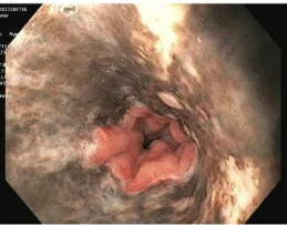
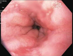
Methods: 43-year-old woman with Type I diabetes mellitus and alcohol use disorder presented with hematemesis. She was found to have diabetic ketoacidosis (DKA) and shock requiring intravenous (IV) pressors. Treatment of DKA with insulin infusion and IV fluids (IVF) was started, in addition to high-dose proton pump inhibitor (PPI) for presumed upper GI bleed. An esophagogastroduodenoscopy (EGD) showed circumferential, black, necrotic-appearing, esophageal mucosa from the mid-esophagus to the gastroesophageal junction (GEJ), and an abrupt transition to normal appearing mucosa (Figure A). Esophageal biopsies revealed acutely inflamed necrotic debris and increased pigmentation, confirming diagnosis of AEN. An EGD repeated one month later showed healing of the esophageal mucosa (Figure B). Discussion: AEN is caused by an ischemic phenomenon seen in low-flow states such as DKA or shock leading to hypoperfusion, tissue injury and mucosal necrosis. The current hypothesized mechanism of injury is believed to be due to a “two hit” hypothesis, where an initial low flow state predisposes the mucosa to a topical injury by gastric acid or pepsin. Although there are no current guidelines, the treatment of AEN is supportive care including IVF, PPIs, bowel rest for 48-72 hours and antibiotics in setting of sepsis. Our patient improved with treatment of DKA and had resolution of necrosis on repeat EGD, which should be considered to rule out stricture formation as this can be seen in approximately 25% of AEN patients. Since AEN has a high mortality of 33%, guidelines for management may be warranted in the future.
Figure: EGD revealed black-colored mucosa in the mid to distal esophagus with transition point of normal mucosa at the GEJ, diagnostic of acute esophageal necrosis.
Figure: Follow up EGD demonstrates healing esophagitis and resolution of AEN.
Disclosures: Nermila Ballmick indicated no relevant financial relationships. Nathaniel Krogel indicated no relevant financial relationships.
Nermila A. Ballmick, MD, Nathaniel Krogel, MD. P4968 - The Black Hole: A Rare Case of Acute Esophageal Necrosis Presenting as an Upper GI Bleed in Diabetic Ketoacidosis, ACG 2025 Annual Scientific Meeting Abstracts. Phoenix, AZ: American College of Gastroenterology.

 photo")