University of Texas Health San Antonio San Antonio, TX
Cody Hu, MD, Andrew Han, MD, Michelle Conde, MD University of Texas Health San Antonio, San Antonio, TX Introduction: Coloniclipomasarebenigntumorscomposedofadiposetissue, which arecommonly asymptomatic.Here,wepresentacaseofcolonicintussusceptioncausedbyagiantcolonic lipomaanditsmanagement.
Case Description/
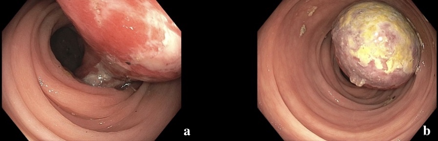
Methods: A59-year-oldfemalewithhypertension and historyoftobaccousepresentedtotheERforacutesharpabdominalpainatbilateral lowerquadrantsoverthepast8days.Thepainwasassociatedwithnauseaandhematochezia. Onarrival,hervitalswerenormal,andphysicalexamwassignificantforthetendernessto palpationintheleftlowerquadrantwithnoreboundingcharacteristic.Labsrevealednormal hemoglobin,lacticacid,andelectrolytes.CTabdomendisclosedaleft-sidedcolonic intussusceptionwitha5cmovoidintraluminalfatdensitymasswhichwasaleadpointand representedacoloniclipoma.SurgeryandGIwereconsultedintheER,andcolonoscopywas urgentlydoneonthefollowingdayforendoscopicreductionandmucosaevaluation(image1). Althoughthecolo-colonicintussusceptionwassuccessfullyreduced,patientdevelopednausea andabdominalpainthedayaftercolonoscopy.RepeatedCTdemonstratedrecurrentcolo-colonicintussusception. Thepatientwassubsequentlytakenforpartialcolectomy(image2). Postoperativecoursewasuneventful,andshewasdischargedfollowingtoleranceofanoral diet. Discussion: Colonic lipomas are often an incidental finding during endoscopic evaluation or imaging studies. The uniform fat density of the lipomas, without any solid component, makes them readily distinguishable on CT and MRI. This feature also underlies the “pillow sign” observed on endoscopy—an indentation on the lesion when pressed with biopsy forceps. It has a 98% specificity, but sensitivity is only limited to 40%. While frequently asymptomatic, giant colonic lipomas—measuring over 4 cm—become symptomatic in approximately 75% of cases. Colonic intussusception is a rare but serious complication of colonic lipomas. In this case, surgery team recommended endoscopic intervention with the hope of achieving therapeutic resection. However, due to the high risk of perforation, absence of pillow sign, and ulceration at the base of stalk, it was not removed with endoscopy. Surgical operation is the first-line treatment option in her circumstance due to the large size. Endoscopic reduction as a bridge to definitive surgical operation to optimize preoperative status highlights the importance of the multidisciplinary approach in managing complicated colonic lipomas.
Figure: Image 1: a) A pedunculated, 5 cm mass with surface ulceration and exudates reaching to the base of the stalk. b) The lesion caused nearly complete obstruction of the lumen, but air and liquid were able to pass after reduction of the intussusception. Pillow sign was negative. No evidence of mucosal ischemia. Intra-procedurally, due to the atypical features, including negative pillow and ulceration, it was not removed with endoscopy.
Figure: Image 2: Surgical specimen demonstrating a large submucosal lipoma with surface ulceration and neighboring colonic tissue.
Disclosures: Cody Hu indicated no relevant financial relationships. Andrew Han indicated no relevant financial relationships. Michelle Conde indicated no relevant financial relationships.
Cody Hu, MD, Andrew Han, MD, Michelle Conde, MD. P4690 - When Benign Becomes Serious: Colonic Lipoma Causing Intussusception, ACG 2025 Annual Scientific Meeting Abstracts. Phoenix, AZ: American College of Gastroenterology.