Tuesday Poster Session
Category: Colon

Rukhsar Rukhsar, MD
Saint Michael's Medical Center, New York Medical College
Parsipanny, NJ
Leiomyoma and GIST (gastrointestinal stroll tumors) are mesenchymal tumors that are typically found incidentally presenting as protruding submucosal lesions. Leiomyoma are generally a benign entity that carry little to no risk of malignancy, GIST carry a higher potential to become malignant. These lesions are typically seen more in the upper gastrointestinal tract rather than lower. On pathology they reveal smooth muscle differentiation and spindle cells.
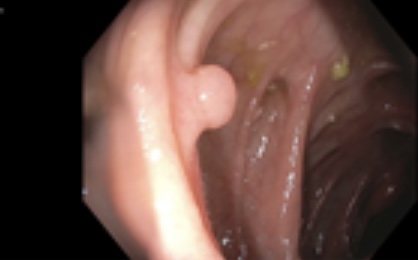
A 53-year-old female with a history of abdominoplasty, endometrial hyperplasia status post hysteroscopy and curettage, and salpingectomy presented for a routine colonoscopy for colorectal cancer screening. She has never had a colonoscopy in the past, and denies any active gastrointestinal symptoms, smoking, alcohol, or substance use. There is no family history of gastrointestinal malignancy. Her physical examination and routine labs were within normal limits. Colonoscopy revealed two semi-pedunculated polyps measuring 6-8 mm in size in the descending colon and ascending colon, which were successfully resected and retrieved with cold snare polypectomy. Histologic examination of ascending revealed a submucosal spindle cell lesion, suspicious for leiomyoma. Subsequent immunohistochemical staining was positive for smooth muscle actin (SMA) and negative for S100, supporting the diagnosis of a submucosal leiomyoma. Interestingly, specimen was also positive for DOG1. Descending colon polyp was diagnosed as a tubular adenoma.
While Leiomyomas and GIST typically look different endoscopically, that is not always the case. DOG-1 positive are generally associated with GIST and its presence in a leiomyoma is very unusual. Studies have demonstrated while rare, on occasion they can be positive if they contain interstitial cells of cajal which can mimic GIST. The pathology does not always match with what is seen endoscopically. Nothing is black and white unfortunately; it is best to work up all polyp appropriately regardless of endoscopic features. Something that seemed so benign can potentially become malignant and can be easily missed.