Staten Island University Hospital Staten Island, NY
Daniel Kalta, DO, MS1, Rajarajeshwari Ramachandran, MD2, Heidi Budke, MD3, Vinaya Gaduputi, MD3 1Staten Island University Hospital, Staten Island, NY; 2Fresno Veterans Affairs Medical Center, Fresno, CA; 3Blanchard Valley Health System, Findlay, OH Introduction: Colonic perineuriomas are uncommon benign mucosal proliferations of mesenchymal cells expressing perineural markers. We are presenting a case of incidental diagnosis of rectal perineurioma during surveillance colonoscopy.
Case Description/
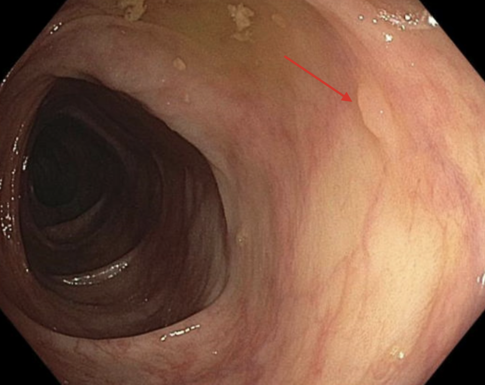
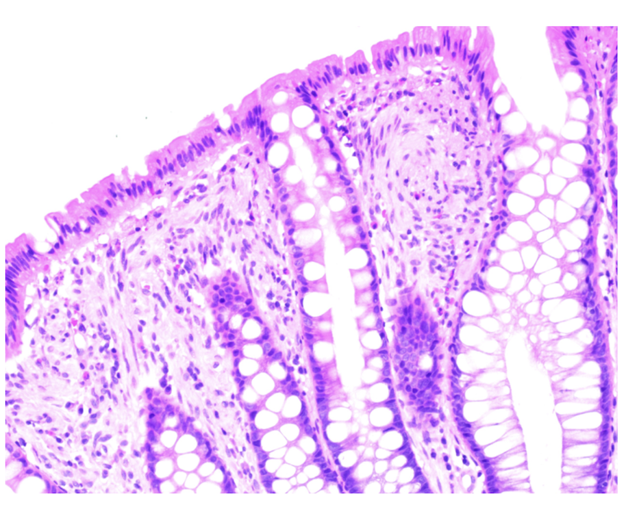
Methods: 55-year-old female without any significant past medical history, presented for surveillance colonoscopy due to prior history of colon adenomas. No family history of colon cancer. No history of nausea, vomiting, abdominal pain, melena, hematochezia, change in bowel habits, and unintentional weight loss. On examination, patient was comfortable, abdomen was soft, non-tender, without organomegaly or ascites. During the colonoscopy, four 2-6 mm sessile polyps (image 1) were removed from the rectosigmoid using cold biopsy forceps and cold snare. Microscopic examination of the polyps demonstrated bland spindle cells in the lamina propria (image 2) and immunostains confirmed the diagnosis of perineurioma. Discussion: Colonic perineurioma is a relatively rare type of benign tumor found in the gastrointestinal tract. The mean age at diagnosis is 60 years, and there is a slight female predominance. 3/4th of the colonic perineuriomas are found in the sigmoid colon or rectum. Histologically, they are composed of spindle cells that fill the lamina propria. Given the benign nature of perineurioma, endoscopic removal is not indicated in asymptomatic patients.

.jpg "Daniel Kalta, DO, MS photo")