Penn State Health Milton S. Hershey Medical Center Hershey, PA
Swapnil Patel, DO, Hadie Razjouyan, MD Penn State Health Milton S. Hershey Medical Center, Hershey, PA Introduction: Radiation is frequently used in the management of abdominal/pelvic malignancies, but it can cause delayed gastrointestinal complications. One late adverse effect is chronic radiation enteritis, characterized by progressive mucosal injury, submucosal fibrosis, and eventual luminal narrowing. The small intestine, particularly the terminal ileum (TI), is especially vulnerable to radiation damage due to its position within the pelvis. Although radiation-induced enteric stenosis is rare, it is clinically significant and often presents with nonspecific symptoms such as abdominal pain and recurrent episodes of partial small bowel obstruction (SBO). Diagnosis is usually difficult given nonspecific imaging findings and lack of surgical findings. We present a case of radiation-induced TI stenosis, diagnosed and treated with endoscopic retrograde enteroscopy.
Case Description/
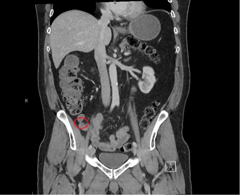
Methods: A 63-year-old male with a history of sigmoid colon cancer treated with surgical resection followed by adjuvant chemoradiation presented with recurrent episodes of partial SBO over ten years. Despite >20 hospital admissions, repeated cross-sectional imaging remained unremarkable. Extensive workup, including negative evaluations for Crohn’s and celiac disease, failed to reveal a definitive cause. Patient underwent two exploratory laparotomies showing no evidence of adhesions, strictures, masses, or mucosal abnormalities. Repeat computed tomography (CT) enterography later revealed evidence of segmental luminal narrowing with hyperemic wall changes near the terminal ileum (Figure 1). The patient subsequently underwent retrograde enteroscopy, which identified severe intraluminal stenosis approximately 20 cm proximal to the ileocecal valve (Figure 2A). Endoscopic balloon dilation was performed up to 10 mm (Figures 2B–C), with biopsies from the stenotic segment showing mild chronic inflammation without evidence of malignancy or granulomatous disease. After dilation, the patient remained symptom-free for months without hospital readmission for partial SBO. Discussion: This case highlights a rare presentation of chronic radiation enteritis causing TI stenosis and recurrent partial SBO. It underscores the importance of considering radiation-induced pathology in patients with a history of abdominal/pelvic radiation and nonspecific obstructive symptoms, even when standard imaging and surgical evaluations are non-diagnostic. Endoscopic evaluation and intervention can serve as both a diagnostic and therapeutic modality in certain cases.
Figure: Figure 1. Computed tomography enterography revealing evidence of segmental luminal narrowing with hyperemic wall changes (red circle) near the terminal ileum
Figure: Figure 2. (A) Terminal illeum stenosis measuring less than 1 cm (in length) x 5 mm (inner diameter) (B) yellow arrow indicating site of dilation with an 8-9-10 mm colonic balloon dilator (C) dilation site showing moderate mucosal disruption and improvement in luminal narrowing
Disclosures: Swapnil Patel indicated no relevant financial relationships. Hadie Razjouyan indicated no relevant financial relationships.
Swapnil Patel, DO, Hadie Razjouyan, MD. P6221 - Radiation-Induced Terminal Ileal Stenosis: Diagnostic and Therapeutic Role of Retrograde Enteroscopy, ACG 2025 Annual Scientific Meeting Abstracts. Phoenix, AZ: American College of Gastroenterology.

.jpg "Swapnil Patel, DO photo")