United Health Services, Wilson Medical Center Johnson City, NY
Fawad Talat, MD1, Khandokar A. Talib, MD2, Abdul Subhan Talpur, MD1, Usama Sakhawat, MD1, Mustafa Sajjad Cheema, MBBS3, Abdul Basit Khan, MD1, Ahmed Shehadah, MD4, Ali Marhaba, MD1 1United Health Services, Wilson Medical Center, Johnson City, NY; 2United Health Services, Wilson Medical Center, Vestal, NY; 3CMH Lahore Medical College and Institute of Dentistry, Lahore, Punjab, Pakistan; 4United Health Services, Wilson Medical Center, Binghamton, NY Introduction: Idiopathic sclerosing mesenteritis is a rare condition with an estimated global prevalence of < 1 %. Radiological imaging of idiopathic sclerosing mesenteritis can have features very similar to carcinoid tumors. We present a case where patient suspected to have carcinoid tumor based on imaging findings was eventually found to have idiopathic sclerosing mesenteritis on histopathology.
Case Description/
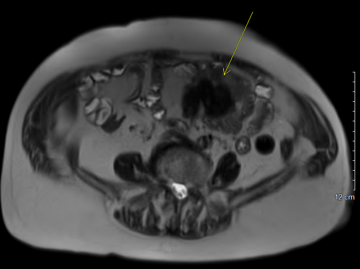
Methods: An 80-year-old female with history of paroxysmal atrial fibrillation, OSA, CHF, recent mitral valve repair, pulmonary hypertension, anxiety, depression and seizure disorder presented with intractable nausea. CT abdomen and pelvis found a partially calcified mesenteric mass in the mid left abdomen, measuring 49 mm. A subsequent MRI abdomen showed a 4.2 x 3.8 cm left abdominal nodular mesenteric mass, bowel wall thickening and desmoplastic reaction, aligning with the diagnosis of a carcinoid tumor.
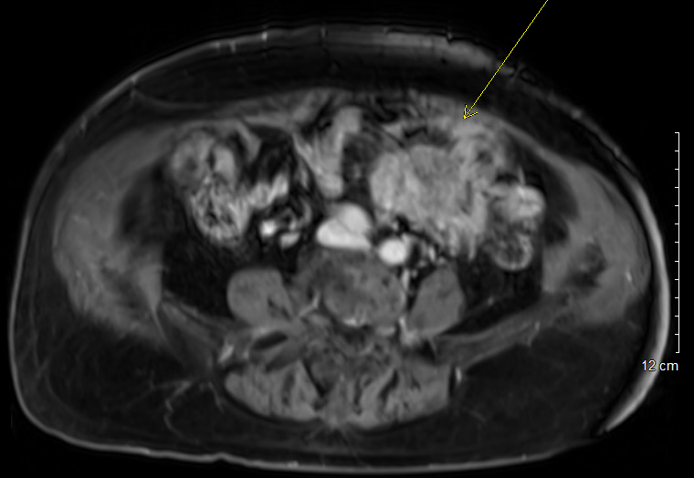
Consequently, the patient underwent exploratory laparotomy with resection of mesenteric mass along with segment of small bowel. Histopathology report of this mesenteric mass revealed no malignant cells but rather showed mesenteric fibrosis, calcification and plasma cell infiltration. Overall, the findings were most consistent with sclerosing mesenteritis. Further workup was carried out to rule out common conditions associated with sclerosing mesenteritis. Autoimmune workup and IgG4 level turned out to be negative. Pan scan also did not show any evidence of malignancy. In this context, a diagnosis of idiopathic sclerosing mesenteritis was established. Discussion: Sclerosing mesenteritis is a chronic inflammatory condition that most commonly affects the mesentery of small intestine and is characterized by inflammation, fibrosis and necrosis of mesentery. It is a rare condition most commonly seen in white men in fifth to seventh decade of life. Although definitive diagnosis requires biopsy, imaging is an important modality to diagnose sclerosing mesenteritis. In this case, MRI initially raised concern for carcinoid tumor based on imaging findings, including a nodular mass, bowel wall thickening, and desmoplastic reaction, features that can be present in both conditions (carcinoid tumor and sclerosing mesenteritis). This case highlights the importance of considering other uncommon diagnosis based on imaging findings.
Figure: Axial T2 FS BLADE image without contrast showing 4.2 x 3.8 cm left abdominal mesenteric mass with features most consistent with carcinoid tumor
Figure: Axial T2 FS BLADE image with IV contrast showing 4.2 x 3.8 cm left abdominal mesenteric mass with features most consistent with carcinoid tumor
Disclosures: Fawad Talat indicated no relevant financial relationships. Khandokar Talib indicated no relevant financial relationships. Abdul Subhan Talpur indicated no relevant financial relationships. Usama Sakhawat indicated no relevant financial relationships. Mustafa Sajjad Cheema indicated no relevant financial relationships. Abdul Basit Khan indicated no relevant financial relationships. Ahmed Shehadah indicated no relevant financial relationships. Ali Marhaba indicated no relevant financial relationships.
Fawad Talat, MD1, Khandokar A. Talib, MD2, Abdul Subhan Talpur, MD1, Usama Sakhawat, MD1, Mustafa Sajjad Cheema, MBBS3, Abdul Basit Khan, MD1, Ahmed Shehadah, MD4, Ali Marhaba, MD1. P6296 - When Inflammation Mimics Malignancy: A Case of Idiopathic Sclerosing Mesenteritis, ACG 2025 Annual Scientific Meeting Abstracts. Phoenix, AZ: American College of Gastroenterology.