Melina Brown, DO1, Nathanial Bartosek, MD2, Nishant Aggarwal, MD3, Fady Banno, MD3, Michael Duffy, MD4 1William Beaumont Hospital, Royal Oak, MI; 2Corewell Health East William Beaumont University Hospital, Royal Oak, MI; 3Corewell Health William Beaumont University Hospital, Royal Oak, MI; 4Corewell Health, Royal Oak, MI Introduction: Small bowel hamartomas are rare and benign lesions that are most commonly associated with hereditary syndromes such as Peutz-Jeghers syndrome. Sporadic small bowel hamartomas are even more rare and often asymptomatic. While these hamartomas do not typically cause significant clinical manifestations, they can occasionally lead to gastrointestinal (GI) complications such as intussusception or bleeding.
Case Description/
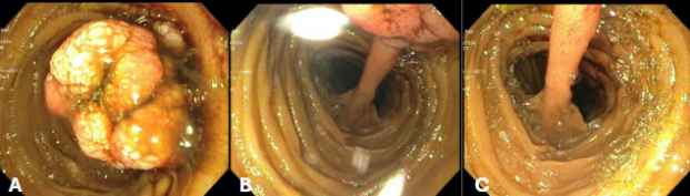
Methods: A 75-year-old gentleman with history of aortic stenosis and Barrett’s esophagus presented after a witnessed syncopal episode at home. He denied any GI symptoms or bleeding. His vitals were stable and physical examination was unremarkable. Initial labs revealed a hemoglobin of 6.3 g/dL (baseline 9 g/dL). Despite multiple blood transfusions, his anemia persisted. Initial endoscopic evaluation including esophagogastroduodenoscopy and colonoscopy were negative. He then underwent video capsule endoscopy (VCE) which revealed a large actively oozing pedunculated jejunal mass. Abdominal CT scan confirmed this small bowel mass. Push enterosocpy (figure 1) showed a large 4 cm multilobulated actively oozing lesion in the jejunum with a long (about 8cm) stalk. The site was tattooed. The patient underwent surgical resection of two benign hamartomatous polyps. Rapid resolution of anemia was noted post operatively after which patient was discharged home in stable condition. On outpatient follow up, he continues to have improving hemoglobin several months later. Discussion: This patient exemplifies a rare instance of a bleeding jejunal hamartoma as a cause of significant iron deficiency anemia. Hamartomatous polyps are usually benign and asymptomatic. After negative EGD and colonoscopy, further investigation with VCE and CT were crucial in identifying the bleeding source. This case demonstrates the importance of including rate and benign lesions in the differential for obscure GI bleeding and shows the importance of advanced imaging techniques in diagnosing potential small bowel pathology when routine endoscopic evaluations are negative.
Figure: Figure 1: Push enteroscopy findings demonstrating 4 cm small bowl hamartoma (A) with attaching 8 cm stalk (B) and stalk origin (C)
Disclosures: Melina Brown indicated no relevant financial relationships. Nathanial Bartosek indicated no relevant financial relationships. Nishant Aggarwal indicated no relevant financial relationships. Fady Banno indicated no relevant financial relationships. Michael Duffy indicated no relevant financial relationships.
Melina Brown, DO1, Nathanial Bartosek, MD2, Nishant Aggarwal, MD3, Fady Banno, MD3, Michael Duffy, MD4. P6294 - Bleeding Small Bowel Hamartoma: A Rare Cause of Iron Deficiency Anemia, ACG 2025 Annual Scientific Meeting Abstracts. Phoenix, AZ: American College of Gastroenterology.