Wayne State University School of Medicine / Detroit Medical Center Detroit, MI
Amro Alhouda, MD1, Mohamed Elhussain, MD2, Abdallah MMEA. Kheshman, MD, MPH2, Mohamed Elbathani, MD2, Ankit MMEA. Hanmandlu, MD2, Bassam Ibrahim, MD3 1Wayne State University School of Medicine / Detroit Medical Center, Detroit, MI; 2Detroit Medical Center - - Detriot, MI, Detroit, MI; 3Detroit Medical Center, Canton, MI Introduction: Gastric glomus tumors (GGTs) are rare mesenchymal neoplasms that are typically benign, with only a handful of cases progressing to metastasis. Malignant transformation and distant spread, especially with vascular invasion, are exceedingly rare and poorly understood. We present the first reported case of a metastatic gastric glomus tumor associated with tumor thrombus extending from the inferior vena cava (IVC) into the right atrium (RA).
Case Description/
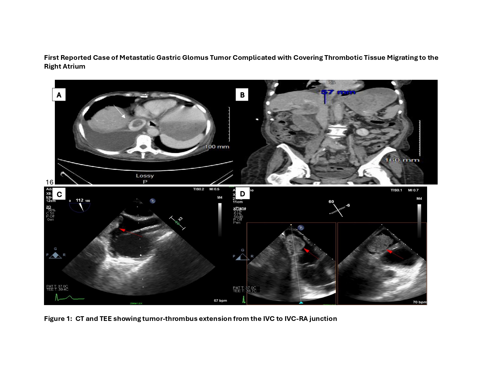
Methods: A 75-year-old woman underwent excision of a gastric mass with partial gastrectomy in 2010 which was thought to be benign. In 2023, she presented with multiple new necrotic metastases throughout the liver and lung which was biopsy-proven to be metastatic GGT. Due to a new finding of tumor thrombus noted in the inferior vena cava (IVC) on CT scan, a TTE was obtained that showed a right atrial mass (1.5 x 2.1 cm). A follow-up TEE with perfusion imaging showed immobile, lobulated, RA mass with heterogenous appearance (2.9 x 2.0 cm) extending from IVC to the IVC-RA junction. Perfusion imaging revealed the mass to be avascular. Discussion: Metastatic spread of GGT is rare, and tumor thrombus involving the IVC-RA junction has not been previously described. Given the patient's significant ascites, frailty, and risk of hemorrhagic or embolic complications, invasive intervention was deemed high-risk. The patient was managed conservatively with anticoagulation for thromboembolic prophylaxis. This case underscores the rare but aggressive behavior of gastric glomus tumors and highlights the importance of long-term surveillance in patients with previously resected GGTs. Gastrointestinal oncologists should maintain a high index of suspicion for vascular involvement in patients presenting with metastatic GGT. Multimodal imaging, including CT and echocardiography, plays a crucial role in detecting tumor thrombus and guiding management in complex gastroenterologic malignancies.
Figure: IVC = Inferior Vena Cava, RA = Right Atrium Axial CT Image of Tumor Thrombus in the IVC Coronal CT Image of Tumor Thrombus in the IVC Bicaval TEE view of Tumor Thrombus obstructing IVC flow RV Inflow (with xPlane) TEE view of Tumor Thrombus in the RA
Disclosures: Amro Alhouda indicated no relevant financial relationships. Mohamed Elhussain indicated no relevant financial relationships. Abdallah Kheshman indicated no relevant financial relationships. Mohamed Elbathani indicated no relevant financial relationships. Ankit Hanmandlu indicated no relevant financial relationships. Bassam Ibrahim indicated no relevant financial relationships.
Amro Alhouda, MD1, Mohamed Elhussain, MD2, Abdallah MMEA. Kheshman, MD, MPH2, Mohamed Elbathani, MD2, Ankit MMEA. Hanmandlu, MD2, Bassam Ibrahim, MD3. P6392 - Metastatic Gastric Glomus Tumor Presenting With Tumor Thrombus Extending to the Right Atrium: A Rare Gastrointestinal Case, ACG 2025 Annual Scientific Meeting Abstracts. Phoenix, AZ: American College of Gastroenterology.