Amr Dokmak, MD1, Camelia Ciobanu, MD1, Madhavi Reddy, MD2, Negar Niknam, MD3 1Brooklyn Hospital Center, Brooklyn, NY; 2The Brooklyn Hospital Center, Brooklyn, NY; 3Icahn school of Medicine at Mount Sinai NYC Health + Hospitals Queens, Gastroenterology Department, Queens, NY Introduction: Gastrointestinal Stromal Tumors (GISTs) are the most common mesenchymal tumors of the gastrointestinal tract. They account for approximately 0.1-3% of all gastrointestinal tumors. GISTs most commonly occur in the stomach (50-60%), followed by the small intestine (30-40%). The average size of a gastric GIST at the time of diagnosis is typically around 5-6 cm, with most tumors discovered incidentally during imaging or endoscopy for unrelated issues. Larger tumors, particularly those exceeding 10 cm, are considered rare and are associated with a higher risk of malignancy and metastasis.
Case Description/
Methods: A 63-year-old man presented to the GI clinic with a 3-month history of progressive abdominal distension and 30-lbs weight loss. A large intra-abdominal mass was seen on physical examination. Laboratory investigations revealed mild anemia. A CT abdomen revealed a 25 cm x 20 cm x 15 cm large, well-defined, heterogeneous mass in the left upper abdomen originating from the stomach and exerting a mass effect on the left hemidiaphragm and spleen. There was displacement of the portal vasculature to the right. No evidence of distant metastasis was noted.
Upper gastrointestinal endoscopy revealed a large deep ulceration in the gastric fundus/body with necrotic tissue, suggestive of a mass invading the lumen. Endoscopic endosonography confirmed the large heterogenous peri-gastric mass with solid and cystic components seen. Biopsy of the gastric mass revealed a mixed GIST with spindle and epitheloid cells present. Tumor cells were positive for CD34, CD117. Ki67 index was 30%. Mitotic index was 18/10 HPF. Patient was referred to oncology and surgery. He was subsequently started on neoadjuvant imatinib. Discussion: This case is notable for the exceptional size of the gastric GIST, measuring 25 cm, which is remarkably larger than the average gastric GIST size of 5-6 cm. While the majority of GISTs are diagnosed at smaller sizes, this case highlights the rarity of such large tumors and underscores the importance of early detection. Larger GISTs have a higher potential for malignancy and metastasis. The management of such tumors typically involves surgical resection, with or without adjuvant therapy, depending on risk factors for recurrence and metastasis.
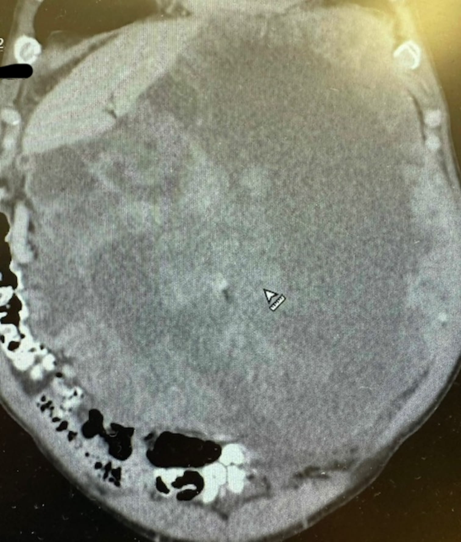
Figure: Figure 1. Abdomen CT demonstrating a massive heterogenous gastric tumor measuring 25 cm x 20 cm x 15 cm with mass effect on spleen and left hemidiaphragm
Disclosures: Amr Dokmak indicated no relevant financial relationships. Camelia Ciobanu indicated no relevant financial relationships. Madhavi Reddy indicated no relevant financial relationships. Negar Niknam indicated no relevant financial relationships.
Amr Dokmak, MD1, Camelia Ciobanu, MD1, Madhavi Reddy, MD2, Negar Niknam, MD3. P6389 - A Case of a Massive Gastrointestinal Stromal Tumor, ACG 2025 Annual Scientific Meeting Abstracts. Phoenix, AZ: American College of Gastroenterology.