Penn State Health Milton S. Hershey Medical Center Hershey, PA
Swapnil Patel, DO1, Raj Aghera, MPH2, Ying Wang, MD1, Ibrahim Yaghnam, MD1, Abraham Mathew, MD1 1Penn State Health Milton S. Hershey Medical Center, Hershey, PA; 2University of North Carolina at Chapel Hill, Wake Forest, NC Introduction: Gastric glomus tumors (GGT) are rare mesenchymal neoplasms, estimated to comprise < 1% of resected gastric lesions. Typically benign, they most commonly arise in the gastric antrum and are composed of modified smooth muscle cells. Clinically, they often present as submucosal masses with nonspecific symptoms, making diagnosis challenging. GGTs are frequently mistaken for gastrointestinal stromal tumors or gastric neuroendocrine tumors (GNET), due to similar clinical and endoscopic features. We present a case of a GGT that was initially suspected to be a GNET, highlighting the diagnostic challenges associated with this rare entity.
Case Description/
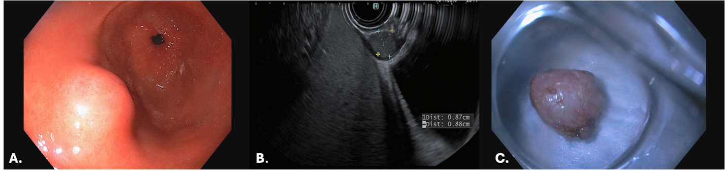
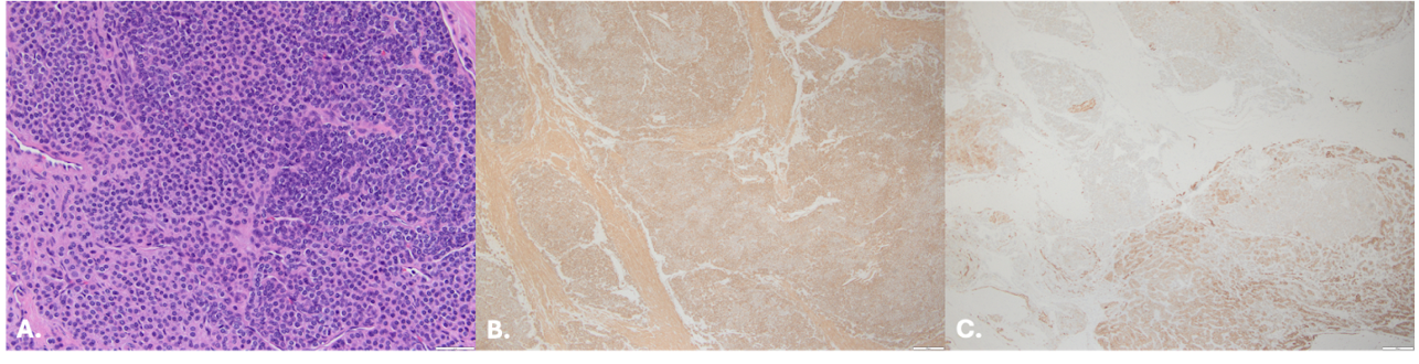
Methods: A 65-year-old male with a history of hypertension, GERD, and anemia presented after an incidentally found gastric antral nodule during a previous esophagogastroduodenoscopy (EGD) (Figure 1A). Endoscopic ultrasound (EUS) revealed an approximately 9 x 9 mm hypoechoic submucosal nodule in the gastric antrum (Figure 1B). Fine needle aspiration (FNA) revealed small uniform cells arranged in organoid nests within the smooth muscle wall, (+) CD56, weak synaptophysin, (-) for chromogranin, DOG1, and CD117, suggesting a well-differentiated grade 1 neuroendocrine tumor. Six months later, the patient underwent endoscopic submucosal dissection (ESD) for management (Figure 1C). Histopathological examination revealed a glomus tumor, characterized by well-circumscribed nodules within the muscularis propria, composed of uniform round cells with a perivascular growth pattern and associated hyalinization (Figure 2A). Immunohistochemistry (IHC) showed diffuse positivity for smooth muscle actin (SMA) (Figure 2B) and focal positivity for synaptophysin (Figure 2C), with (-) staining for cytokeratin AE1/3 and chromogranin. Discussion: This case highlights the diagnostic challenges of GGTs, which often mimic other submucosal lesions due to overlapping clinical, endoscopic, and IHC features. Some GGTs strongly express synaptophysin, resembling GNETs. Unlike GNETs, GGTs lack keratin expression and are centered in the muscularis propria. In this patient, initial FNA biopsy suggested a GNET, however final diagnosis of GGT was achieved only after ESD and comprehensive histopathological evaluation. While EUS-guided FNA can aid diagnosis, its yield may be limited by small sample size, making ESD with full-thickness biopsy more reliable. Clinicians should consider GGTs in the differential diagnosis of submucosal tumors, particularly cases with focal synaptophysin reactivity.
Figure: Figure 1. (A) EGD shows a single submucosal tumor in the gastric antrum (B) EUS revealing an oval hypoechoic intramural (subepithelial) lesion approximately 9 x 9 mm (C) resected tumor status post ESD
Figure: Figure 2. (A) Histology of resected gastric antral mass with H&E staining (20x) shows multiple neoplastic nodules in the gastric muscularis propria, composed of uniform round cells with lightly eosinophilic to clear cytoplasm and a perivascular growth pattern. The tumor cells are strongly and diffusely positive for SMA 4x (B), focally positive for synaptophysin 4x (C)
Disclosures: Swapnil Patel indicated no relevant financial relationships. Raj Aghera indicated no relevant financial relationships. Ying Wang indicated no relevant financial relationships. Ibrahim Yaghnam indicated no relevant financial relationships. Abraham Mathew indicated no relevant financial relationships.
Swapnil Patel, DO1, Raj Aghera, MPH2, Ying Wang, MD1, Ibrahim Yaghnam, MD1, Abraham Mathew, MD1. P6358 - A Gastric Masquerade: Glomus Tumor Mimicking Gastric Neuroendocrine Neoplasm, ACG 2025 Annual Scientific Meeting Abstracts. Phoenix, AZ: American College of Gastroenterology.


.jpg "Swapnil Patel, DO photo")