Rawia Aburumman, MD1, Lena Kawji, MD2, Sudha Pandit, MD2, Kshitij Arora, MD3 1Louisiana State University, Spring, TX; 2Louisiana State University, Shreveport, LA; 3LSU Health, Shreveport, LA Introduction: Pseudomelanosis is a rare endoscopic finding, most commonly involving the duodenum, and characterized by dark pigmentation of the mucosa due to deposition of substances such as iron, melanin, or lipofuscin. Gastric involvement is exceedingly rare, with few cases reported. We present a unique case of gastric pseudomelanosis in a patient whose upper endoscopy one year earlier had shown normal mucosa.
Case Description/
Methods: A 78-year-old female with end-stage renal disease, coronary artery disease, type 2 diabetes, and COPD presented with shortness of breath and dysphagia. Her medications included carvedilol, hydralazine, and ferrous sulfate. One year prior, she was found to have normocytic normochromic anemia (Hgb 6.5 g/dL) and underwent EGD, which showed normal mucosa. She received blood transfusions but continued to take iron supplements daily for the following year. On repeat presentation, she had dysphagia and EGD revealed diffuse mild gastritis with dark mucosal pigmentation in the stomach and duodenum. Gastric biopsy stained with Perl’s iron confirmed crystalline iron deposits, consistent with pseudomelanosis. Discussion: Pseudomelanosis of the stomach is an uncommon, benign condition often associated with chronic renal failure, diabetes, aging, and prolonged iron supplementation. Its pathogenesis may involve impaired mucosal absorption of pigment-forming substances. Recognition of this entity is important to avoid misdiagnosis and unnecessary interventions. Endoscopic findings may mimic serious pathology such as metastatic melanoma; however, biopsy with iron staining is diagnostic. This case highlights the evolving nature of mucosal pigmentation and the need for histological confirmation in atypical findings.
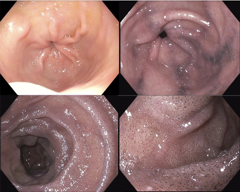
Figure: Upper left image showing normal mucosa during patient's first EGD, and remaining images showing hyper-pigmented mucosa on EGD 1 year later.
Figure: Gastric tissue biopsy stained with Prussian blue (Perl’s iron) stain, which is used to detect iron deposits in tissue.
Disclosures: Rawia Aburumman indicated no relevant financial relationships. Lena Kawji indicated no relevant financial relationships. Sudha Pandit: Medtronic – Product feedback. Kshitij Arora indicated no relevant financial relationships.
Rawia Aburumman, MD1, Lena Kawji, MD2, Sudha Pandit, MD2, Kshitij Arora, MD3. P6355 - Pseudomelanosis of the Stomach: A Rare Gastric Presentation of a Common Duodenal Finding, ACG 2025 Annual Scientific Meeting Abstracts. Phoenix, AZ: American College of Gastroenterology.