P6344 - Gastric Schwannoma versus Gastrointestinal Stromal Tumor: A Case Report and the Future of Artificial Intelligence in Differentiating Subepithelial Gastric Lesions
Mohamed Alharami, MD1, Omar Alwan, MD2, Frhaan Zahrawi, MBBCh3, Omar Khattab, MD4, Zachary Neubert, DO5 1Henry Ford Warren, Warren, MI; 2Hamilton Medical Center, Dalton, GA; 3Franciscan Health Olympia Fields, Olympia Fields, IL; 4Kettering Health Netwrok, Kettering, OH; 5Halifax Medical Center, Daytona Beach, FL, Port Orange, FL Introduction: Gastric schwannoma (GS) is a rare mesenchymal tumor comprising about 0.2% of all gastric neoplasms. Clinical and radiologic presentations often mimic more common gastric subepithelial lesions (SELs), particularly gastrointestinal stromal tumors (GISTs). Although mostly benign, diagnostic uncertainty frequently leads to invasive procedures such as biopsy or surgical resection. Immunohistochemistry (IHC) showing S-100 protein with the absence of other mesenchymal markers remains the standard method to differentiate GS from other SELs like GIST. As artificial intelligence (AI) advances in medical imaging, it holds promise for improving the non-invasive differentiation of SELs, possibly reducing the need for invasive procedures in cases like GS. We present a case of GS along with a brief literature review, emphasizing current diagnostic challenges and the prospective role of AI-assisted imaging.
Case Description/
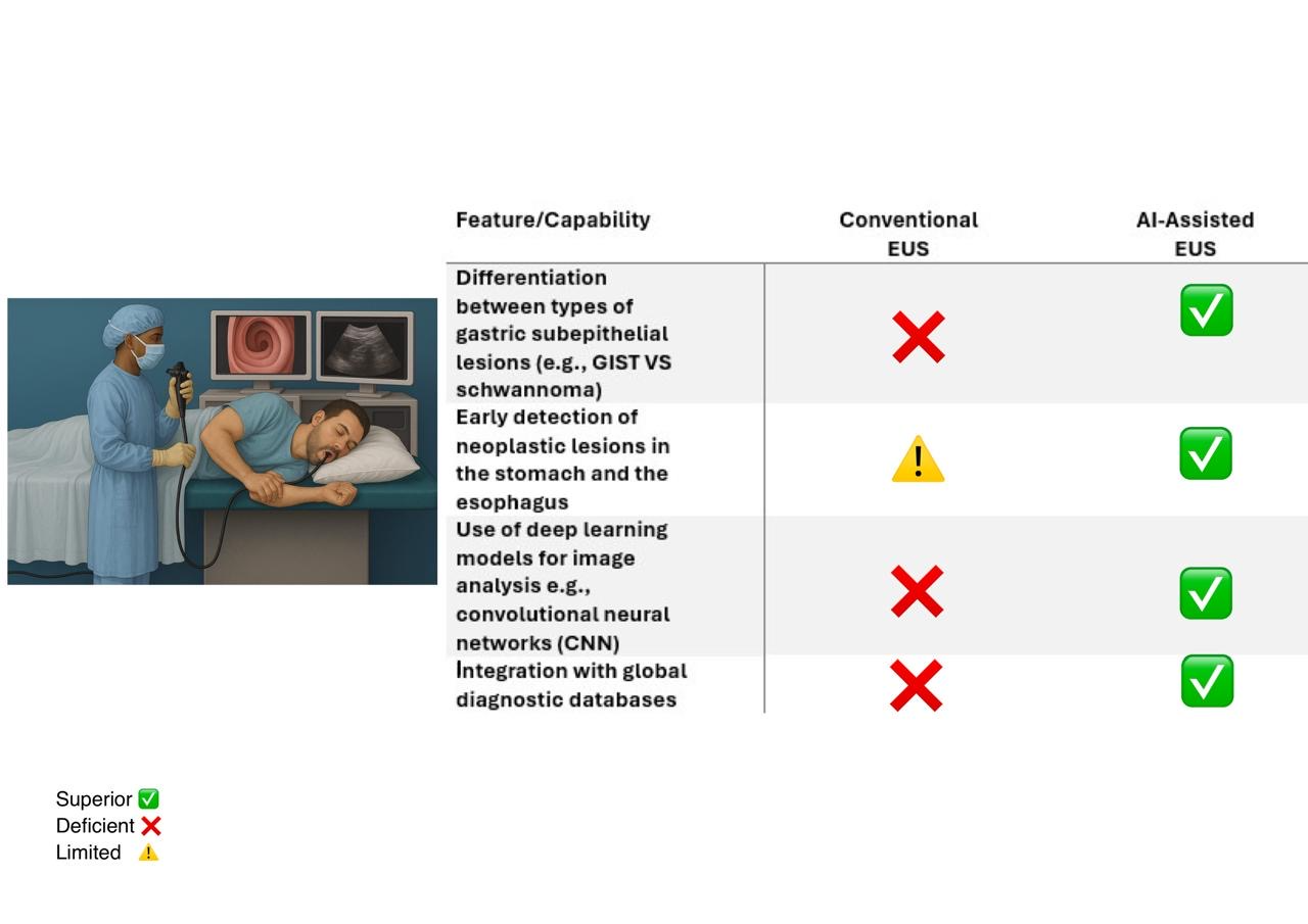
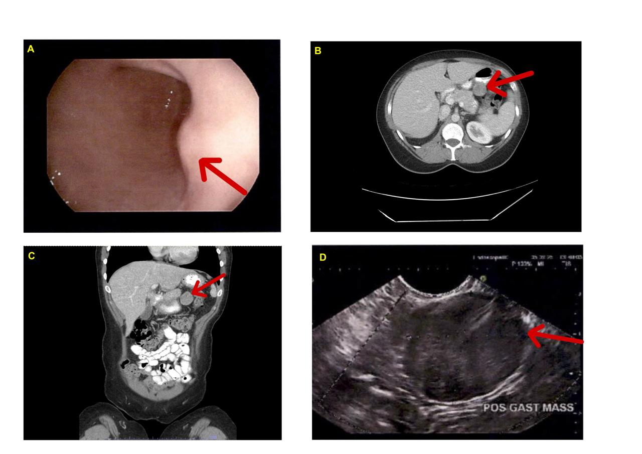
Methods: A 37-year-old woman presented with several weeks of worsening epigastric pain and nausea. Dicyclomine and a proton pump inhibitor (PPI) were started, but with no relief of symptoms. A CT scan of the abdomen showed a 3 cm mass arising from the posterior gastric wall, raising concern for possible GIST (Figure 1). Subsequent esophagogastroduodenoscopy (EGD) with endoscopic ultrasound-guided fine-needle aspiration (EUS-FNA) demonstrated a submucosal bulge with a well-defined hypoechoic mass originating from the gastric muscularis propria (Figure 1). Biopsy revealed a spindle cell neoplasm positive for S-100 and negative for other mesenchymal markers including CD34, CD117, desmin, and Ki-67. Additionally, negative IHC for HMB-45, PRAME, and Melan-A favored a diagnosis of gastric schwannoma over melanoma. The patient was referred to a specialized surgical center for definitive management. Discussion: GS shares imaging features with GISTs, making preoperative diagnosis challenging. Direct tissue biopsy with IHC remains the diagnostic gold standard. Surgical resection is curative, and unlike GISTs, GS does not require systemic therapy. Due to overlapping imaging features among gastric SELs, AI has recently demonstrated promise in improving imaging interpretation. Some AI systems can now perform real-time classification and distinguish multiple lesion types using subtle imaging patterns not readily detected by human readers (Figure 2). While still under development, AI-assisted imaging may soon play a central role in aiding non-invasive differentiation between GS and other SELs.
Figure: Figure 1. (A) Esophagogastroduodenoscopy (EGD) revealing a submucosal bulge in the posterior gastric body (red arrow). (B) Axial abdominal CT scan showing a 3 cm exophytic mass arising from the posterior wall of the gastric body (red arrow). (C) Coronal CT image further delineating the exophytic nature and origin of the mass (red arrow). (D) Endoscopic ultrasound (EUS) identifying a well-defined, hypoechoic mass originating from the muscularis propria layer (red arrow).
Figure: Figure 2. Comparison of conventional EUS and AI-assisted EUS in key diagnostic capabilities, highlighting the superior performance of AI-assisted imaging in lesion differentiation, early detection, and data integration. *EUS = Endoscopic Ultrasound
Disclosures: Mohamed Alharami indicated no relevant financial relationships. Omar Alwan indicated no relevant financial relationships. Frhaan Zahrawi indicated no relevant financial relationships. Omar Khattab indicated no relevant financial relationships. Zachary Neubert indicated no relevant financial relationships.
Mohamed Alharami, MD1, Omar Alwan, MD2, Frhaan Zahrawi, MBBCh3, Omar Khattab, MD4, Zachary Neubert, DO5. P6344 - Gastric Schwannoma versus Gastrointestinal Stromal Tumor: A Case Report and the Future of Artificial Intelligence in Differentiating Subepithelial Gastric Lesions, ACG 2025 Annual Scientific Meeting Abstracts. Phoenix, AZ: American College of Gastroenterology.