
Monday Poster Session
Category: Colon
Hareem Tahir, MD
University of South Dakota Sanford School of Medicine
Sioux Falls, SD
Patient is a 77-year-old male with a past medical history of UC who presented with abdominal pain. Labs revealed transaminitis with significant conjugated hyperbilirubinemia. Imaging showed a mass in the liver concerning for an abscess. This was initially treated with broad-spectrum antibiotics and drain placement with no clinical improvement. The decision was made to perform a liver biopsy, which revealed carcinoma with squamous cell differentiation. Tempus tumor of origin testing was inconclusive. Later, the patient underwent a colonoscopy and esophagogastroduodenoscopy (EGD) to find a potential primary site for malignancy. EGD was negative, and colonoscopy revealed a mass at the hepatic flexure of the colon with pathology consistent with SCC. PET scan (Positron Emission Tomography scan) did not reveal any other sites suspicious for primary malignancy. Hence colon was identified to be the primary site with extension to the liver. He was not a surgical candidate and was initiated on FOLFOX chemotherapy.
Discussion:
Primary SCC of the colon is a rare malignancy (less than 0.1% of all colonic malignancies). Its pathogenesis remains poorly understood. UC is a recognized risk factor for colonic adenocarcinoma, but the association with SCC colon is not well established.
Diagnosis can be difficult due to nonspecific symptoms and radiographic features indistinguishable from other common colon malignancies. In this case, the hepatic lesion was misinterpreted as an abscess, which delayed diagnosis. The identification of SCC on liver and colon biopsy, combined with negative EGD and mass on colonoscopy extending into the liver, supported the colon as the primary site. Given aggressive behavior and presentation with advanced disease, the prognosis is typically poor. There are no consensus guidelines for management, but surgery is considered the mainstay. In nonsurgical cases, platinum-based chemotherapy regimens are employed, although outcomes remain suboptimal.
This case highlights diagnostic and therapeutic challenges in a rare colonic malignancy with need for more evidence to guide treatment.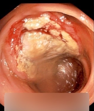
Figure: Figure 1: Mass seen during colonoscopy, later found to be Squamous cell carcinoma.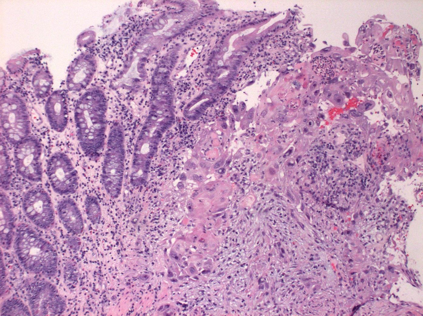
Figure: Figure 2: Microscopic examination of colonic mass showing glandular colonic mucosa next to squamous cell carcinoma.
Disclosures:
Hareem Tahir indicated no relevant financial relationships.
Muhammad Zahid Anwar indicated no relevant financial relationships.
Rafia Irfan Waheed indicated no relevant financial relationships.
Erin Quist indicated no relevant financial relationships.
Hareem Tahir, MD1, Muhammad Zahid Anwar, MBBS2, Rafia Irfan Waheed, MD1, Erin Quist, MD3. P2488 - A Rare Case of Primary Squamous Cell Carcinoma of the Colon in a Patient With Ulcerative Colitis, ACG 2025 Annual Scientific Meeting Abstracts. Phoenix, AZ: American College of Gastroenterology.