University of Utah School of Medicine Salt Lake City, UT
Benjamin Gow-Lee, MD1, Jessica Schmitt, MD1, John Erikson Yap, MD, MBA, FACG2 1University of Utah School of Medicine, Salt Lake City, UT; 2University of Utah Health, Salt Lake City, UT Introduction: While adenocarcinoma accounts for the overwhelming majority of colorectal malignancies, it is crucial to consider alternative and less common pathologies when evaluating colonic masses. These include lymphomas, carcinoid tumors, neuroendocrine tumors, and squamous cell carcinoma—each with distinct pathophysiology, clinical progression, and treatment strategies. Failure to consider these differential diagnoses can delay appropriate therapy and impact patient outcomes. We present a rare case of mantle cell lymphoma (MCL), an uncommon gastrointestinal lymphoma, presenting as an isolated cecal mass that was initially suspected to be adenocarcinoma based on endoscopic appearance.
Case Description/
Methods: A 70-year-old man with a history of coronary artery disease and longstanding iron deficiency anemia was referred for evaluation of abdominal pain, worsening anemia, and unintentional weight loss. As part of his workup, both esophagogastroduodenoscopy (EGD) and colonoscopy were performed. The EGD was grossly unremarkable. However, colonoscopy revealed a large, fungating, and partially obstructing mass within the cecum extending into the ileocecal valve. Biopsies of the lesion confirmed the unexpected diagnosis of mantle cell lymphoma. Further staging with PET-CT revealed intussusception at the ileocecal junction and mesenteric lymphadenopathy, but no evidence of distant metastatic disease. Given the lack of obstructive symptoms, surgical intervention was deferred. The patient was managed with rituximab, lenalidomide, and localized cecal radiation and showed favorable clinical response. Discussion: MCL is a rare subtype of B-cell non-Hodgkin lymphoma. While it typically presents with generalized lymphadenopathy, gastrointestinal involvement can occur in up to one-third of patients. GI involvement often presents as multiple lymphomatous polyps but can also appear as isolated mass lesions. This case highlights the importance of maintaining a broad differential when evaluating colonic lesions. Early tissue diagnosis is essential, as the management of lymphomas differs significantly from adenocarcinomas and relies primarily on systemic rather than surgical therapy.
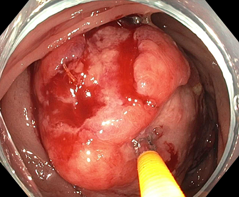
Figure: Cecal mass found to be mantle cell lymphoma.
Figure: Hematoxylin and Eosin (H&E) stain demonstrating a medium-power view of a cecal mass biopsy. A submucosal lymphoid infiltrate (left arrow) is identified below normal appearing colonic mucosa (right arrow). Image was captured with a 20x objective.
Disclosures: Benjamin Gow-Lee indicated no relevant financial relationships. Jessica Schmitt indicated no relevant financial relationships. John Erikson Yap: Phathom Pharmaceutical – Speakers Bureau. Steris – Consultant.
Benjamin Gow-Lee, MD1, Jessica Schmitt, MD1, John Erikson Yap, MD, MBA, FACG2. P2579 - Not All Colon Masses Are Adenocarcinoma: An Unusual Case of Mantle Cell Lymphoma, ACG 2025 Annual Scientific Meeting Abstracts. Phoenix, AZ: American College of Gastroenterology.

.jpg "Benjamin Gow-Lee, MD (he/him/his) photo")