Sevag Hamamah, DO, Nupur Savalia, MD, Faizi Hai, MD Scripps Mercy Hospital, San Diego, CA Introduction: Proctitis, or inflammation of the rectal mucosa, stems from various etiologies including inflammatory bowel disease, radiation, ischemia, or infection. Sexually transmitted infection (STI) from Neisseria gonorrhoeae or Chlamydia trachomatis is the most common infectious cause of proctitis in young patients. Risk factors include previous STI, high-risk sexual behaviors, men who have sex with men and immunocompromised status. Chlamydia trachomatis has lymphogranuloma venereum (LGV) serovars (L1, L2, L3) that may present as painless ulcers or painful hemorrhagic proctitis and can progress to invasive disease. However, presentation as a rectal mass with lymphadenopathy is rare and poses diagnostic challenges. We present a rare case of proctitis secondary to LGV and Neisseria gonorrhoeae coinfection in an immunocompromised individual presenting as a rectal mass.
Case Description/
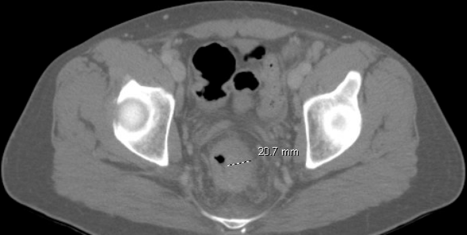
Methods: A 37-year-old man with history of neurosyphilis and human immunodeficiency virus (CD4 count: 350 cells/mm3), noncompliant with antiviral treatment, presented with two months of intermittent rectal pain and bleeding. Computed tomography (CT) abdomen and pelvis showed a rectal annular mass extending 2cm in diameter with increased rectal wall thickness and possible extension through the rectal wall. Numerous lymph nodes in the mesorectal fascia, pelvic sidewalls and bilateral inguinal regions were also identified. Colonoscopy was performed with findings of mild erythema in the descending colon, moderate erythema and mild superficial ulceration in the sigmoid colon, and diffuse edema and erythema with 2-3cm coalescing deep ulcers in the rectum without active bleeding. Rectal, sigmoid colon, and descending colon biopsies demonstrated active colitis with crypt abscess formation, focal erosion, and focal granulation tissue. Rectal swab was positive for Chlamydia trachomatis and Neisseria gonorrhoeae. Patient received a 2 gram one-time dose of ceftriaxone and doxycycline 100mg twice daily for 28 days, with gradual improvement in symptoms. Discussion: This case illustrates LGV proctitis with gonorrhea coinfection mimicking a locally invasive rectal mass, driven by inflammatory changes and regional lymphadenopathy. Despite this unusual presentation, it is important to consider infectious etiologies in high-risk patients, as early diagnosis and treatment is important in preventing LGV-related complications including deep mucosal ulcerations, rectal strictures, perirectal abscesses, fistulas or even disseminated disease.
Figure: Figure 1. Computed Tomography of the abdomen and pelvis showing diffuse rectal wall thickening with a rectal mass. The rectal wall is irregular in appearance and thickened to 2cm in diameter.
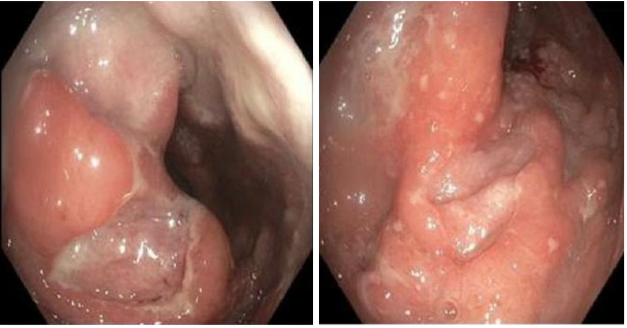
Figure: Figure 2. Colonoscopy images demonstrating diffuse edema and erythema with large (2-3cm) coalescing deep ulcerations without active bleeding.
Disclosures: Sevag Hamamah indicated no relevant financial relationships. Nupur Savalia indicated no relevant financial relationships. Faizi Hai indicated no relevant financial relationships.
Sevag Hamamah, DO, Nupur Savalia, MD, Faizi Hai, MD. P2573 - Proctitis Secondary to Lymphogranuloma Venereum and Neisseria Gonorrhoeae Coinfection Presenting as Rectal Mass, ACG 2025 Annual Scientific Meeting Abstracts. Phoenix, AZ: American College of Gastroenterology.