Alaska Digestive and Liver Disease, LLC Eagle River, AK
Jack E. Molloy, 1, Austin Nelson, MD2 1Alaska Digestive and Liver Disease, LLC, Anchorage, AK; 2Alaska Digestive and Liver Disease, Anchorage, AK Introduction: We present a rare case of Basal Pattern Squamous Dysplasia present throughout the esophagus. Squamous dysplasia without carcinoma typically presents as a discrete lesion in the esophagus. This case represents a unique occurrence in which squamous epithelium has developed into dysplasia throughout the entire esophagus. Dysplasia was identified at the base of the epithelium only. This case offers insight into the natural history of this uncommon diagnosis and the histologic outcomes following ablative treatments.
Case Description/
Methods: The patient is a 67-year-old woman suffering from over a decade of GERD. In 2017, the patient was diagnosed by biopsy with only mild inflammation of the upper and lower thirds of the esophagus without atypia or dysplasia. In 2023, she returned with worsening symptoms of reflux and solid food dysphagia. Endoscopy revealed a normal appearance under white light and narrow-band imaging with high-definition resolution. Biopsies demonstrated esophageal squamous mucosa with basal zone squamous atypia in both the upper and lower thirds of the esophagus. These findings were consistent with the rarely observed diagnosis of basal zone squamous dysplasia. Specimens were sent for expert consultation. Ki-67 showed increased proliferation limited to the basal portion of the mucosa. Final pathologic diagnosis was Basal Pattern Squamous Dysplasia throughout the esophagus. Immunostains performed on the samples throughout the esophagus at 2-4cm intervals show that they are diffuse positive for DSG-3, focally positive for p40, while being negative for CDX2 and SATB2. P53 testing shows wild-type staining. The pathologist expressed concern for intraepithelial spread of either high-grade squamous dysplasia or an unsampled carcinoma.
Radiofrequency ablation was performed to eliminate the dysplasia. Despite multiple rounds of RFA and one round of cryoablation, the Basal Zone Squamous Dysplasia persists. Further treatment options are being considered. Discussion: Basal Pattern Squamous Dysplasia is a rare entity. It has been described throughout the esophagus without obvious risk factors. Best practices and standards of care for the treatment of this diagnosis do not exist. The patient declined surgical esophagectomy. Despite numerous rounds of RFA and cryoablation, the pathologic findings persist. Unfortunately, the patient has suffered from esophageal strictures associated with the treatments. The definitive therapy for this uncommon finding remains unknown.
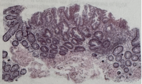
Figure: Pathology in 2017 showing unremarkable squamous mucosa of the esophagus.
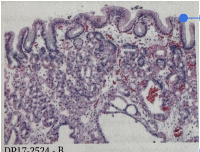
Figure: Pathology in 2023 showing Basal Pattern Squamous Dysplasia
Disclosures: Jack Molloy indicated no relevant financial relationships. Austin Nelson indicated no relevant financial relationships.
Jack E. Molloy, 1, Austin Nelson, MD2. P2820 - Basal Pattern Squamous Dysplasia of the Esophagus, ACG 2025 Annual Scientific Meeting Abstracts. Phoenix, AZ: American College of Gastroenterology.

 photo")