Gagan Midathala, BS1, Fernando Cordero-Baez, MD1, Melissa O'Neal, BS, PharmD2, Alexander Coughlin, PharmD2, Jose Montero, MD1 1USF Health Morsani College of Medicine, Tampa, FL; 2Tampa General Hospital / University of South Florida, Tampa, FL Introduction: Cutaneous manifestations of IBD, such as pyoderma gangrenosum (PG), can precede gastrointestinal (GI) symptoms, and mimic infection or vasculitis, delaying diagnosis. We present a case of a young male whose painful skin ulcers, initially thought to be infectious or autoimmune in origin, led to a new diagnosis of Crohn’s disease (CD).
Case Description/
Methods: A 27-year-old uninsured Mexican male presented to the emergency department with painful ulcers on his thighs, groin, and right arm, along with fever, chills, and diarrhea. The ulcers began 1.5 weeks prior as small pustules that rapidly expanded. He denied recent travel, new medications, or known medical conditions; but, admitted chronic diarrhea, recurrent aphthous ulcers, and similar skin lesions three years earlier. His mother and sister have lupus.
On admission, he was febrile and tachycardic. Skin exam revealed well-circumscribed, tender ulcers with erythematous borders in different stages. Tests showed leukocytosis, normocytic anemia, hypoalbuminemia, hyponatremia, and transaminitis. Broad-spectrum antibiotics were initiated for presumed sepsis secondary to cellulitis.
Dermatology, infectious diseases (ID), and rheumatology were consulted. Blood cultures, ANA, HIV, viral hepatitis panels, AFB culture, and stool studies were negative. CRP and ESR were elevated. Skin biopsy showed superficial and mid-dermal perivascular and periadnexal dermatitis with microabscesses. The wound culture grew Pseudomonas aeruginosa, supporting an initial infectious etiology. Patient was prescribed cefepime with mild improvement.
However, ID remained concerned for a non-infectious primary etiology and gastroenterology was consulted. Colonoscopy revealed severely erythematous and ulcerated mucosa consistent with Crohn's disease. Dermatology then suspected PG as the cause of the skin lesions. Systemic and topical corticosteroids resulted in rapid improvement of both GI and cutaneous symptoms. Discussion: This case underscores the importance of an interdisciplinary approach to a syndromic diagnosis for complex cutaneous ulcers. PG can mimic other conditions delaying diagnosis and treatment of underlying IBD. This patient’s limited access to healthcare as an uninsured Hispanic male emphasizes the impact of health disparities. Collaboration among specialties led to a correct diagnosis and favorable outcome.
AI assistance used in writing portions of this summary.
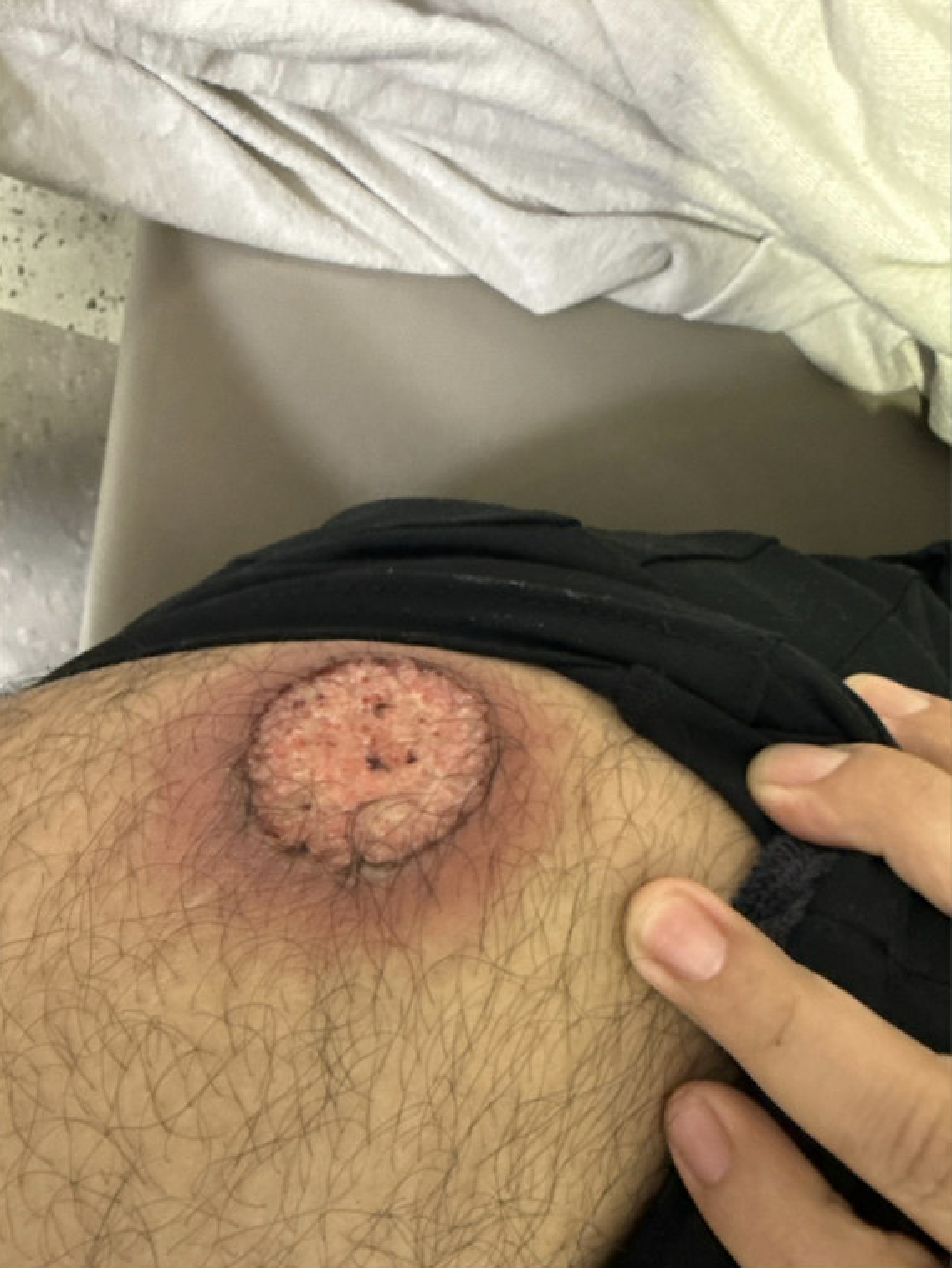
Figure: Lesion with surrounding erythema
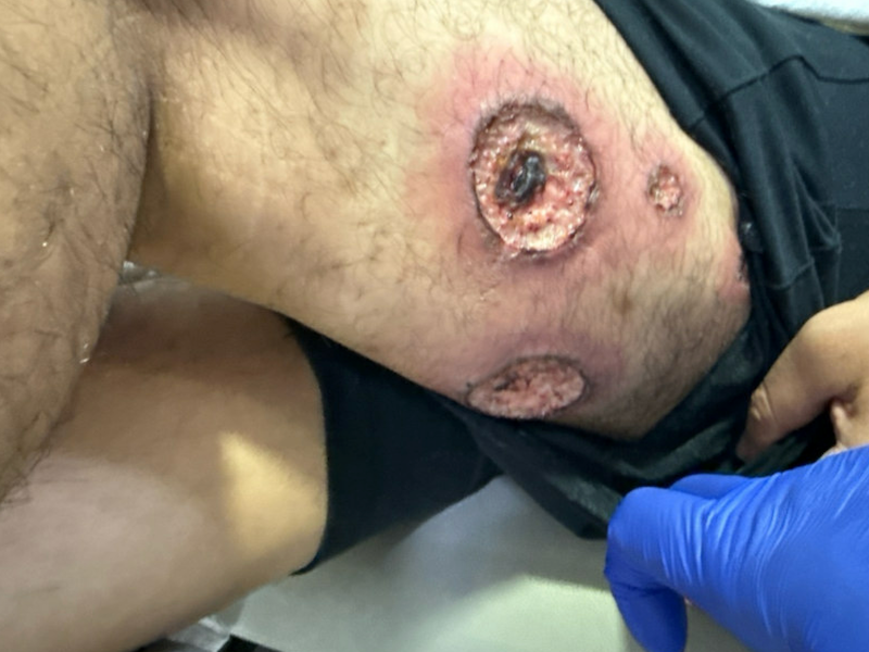
Figure: Left thigh lesions
Disclosures: Gagan Midathala indicated no relevant financial relationships. Fernando Cordero-Baez indicated no relevant financial relationships. Melissa O'Neal indicated no relevant financial relationships. Alexander Coughlin indicated no relevant financial relationships. Jose Montero indicated no relevant financial relationships.
Gagan Midathala, BS1, Fernando Cordero-Baez, MD1, Melissa O'Neal, BS, PharmD2, Alexander Coughlin, PharmD2, Jose Montero, MD1. P3370 - Cutaneous Ulcers as the First Manifestation of Inflammatory Bowel Disease in a Young Male: An Interdisciplinary Diagnostic Challenge, ACG 2025 Annual Scientific Meeting Abstracts. Phoenix, AZ: American College of Gastroenterology.

 photo")