King Faisal Specialist Hospital and Research Centre Ar Riyad, Ar Riyad, Saudi Arabia
Naryman Albadawi, MD1, Mohammed Alayed, MD1, Nawaf Bin Mugren, MD2, Ahmad Bazarbashi, MD3, Aymen Almuhaidb, MD1 1King Faisal Specialist Hospital and Research Centre, Riyadh, Ar Riyad, Saudi Arabia; 2King Faisal Specialist Hospital and Research Centre, Ar Riyad, Ar Riyad, Saudi Arabia; 3Washington University School of Medicine in St. Louis / Barnes-Jewish Hospital, St. Louis, WA Introduction: Fibrosarcoma is a rare malignant tumor of spindle-shaped fibroblasts, typically found in deep soft tissues of the extremities and trunk. Retroperitoneal Fibrosarcoma with osseous metastases is extremely rare and can be a challenging diagnosis. This report discusses a rare case of retroperitoneal metastatic fibrosarcoma mimicking a pancreas mass and identified via endoscopic ultrasound (EUS) guided biopsy.
Case Description/
Methods: A 48-year-old man with no significant medical history presented with one year history of chronic right shoulder pain, mild abdominal discomfort and unexplained weight loss. Magnetic resonance imaging (MRI) of his shoulder showed questionable malignancy due to bone marrow lesions. A subsequent Positron Emission Tomography (PET) scan revealed a FDG-avid retroperitoneal peripancreatic mass and a suspicious lesion in the right humeral head, concerning for metastasis.
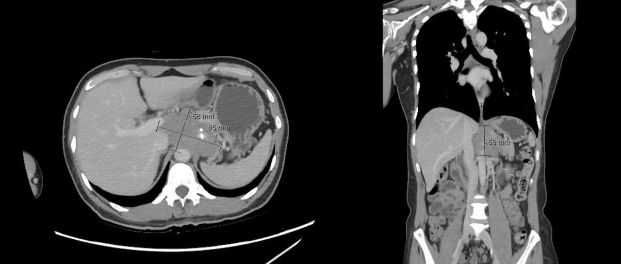
Further imaging via computed tomography (CT) of the abdomen confirmed an ill-defined 5.7 × 8.6 × 6 cm heterogeneous mass in the retroperitoneal area, suspicious for pancreatic adenocarcinoma, affecting surrounding organs and vasculature [figure 1]. EUS revealing a hypoechoic mass greater than 5 cm in the peri-pancreas region. An EUS-guided biopsy of this mass showed spindle cell neoplasm and a CT-guided bone biopsy of the humeral lesion showed spindle cells with sclerosis indicated sclerosing epithelioid fibrosarcoma through specific immunohistochemical staining.
The case was reviewed in a tumor board and confirmed retroperitoneal fibrosarcoma, leading to the initiation of treatment with pazopanib at 800 mg. After two months of treatment, a follow-up PET scan revealed metabolic improvement in the retroperitoneal mass, but new metastatic lung nodules had developed. Discussion: This case highlights the diagnostic utility of EUS guided biopsy in evaluating retroperitoneal masses, which may often mimic pancreatic adenocarcinoma. Clinicians should consider rare tumors like sclerosing epithelioid fibrosarcoma in the differential diagnosis of retroperitoneal masses especially when associated with atypical metastatic pattern.
Figure: Figure 1: transverse and coronal views of cross-sectional imaging revealing large retroperitoneal peri-pancreas mass.
Disclosures: Naryman Albadawi indicated no relevant financial relationships. Mohammed Alayed indicated no relevant financial relationships. Nawaf Bin Mugren indicated no relevant financial relationships. Ahmad Bazarbashi indicated no relevant financial relationships. Aymen Almuhaidb indicated no relevant financial relationships.
Naryman Albadawi, MD1, Mohammed Alayed, MD1, Nawaf Bin Mugren, MD2, Ahmad Bazarbashi, MD3, Aymen Almuhaidb, MD1. P3571 - Beyond the Pancreas: Retroperitoneal Sclerosing Epithelioid Fibrosarcoma Presenting as a Pancreatic Mass With Osseous Metastases, ACG 2025 Annual Scientific Meeting Abstracts. Phoenix, AZ: American College of Gastroenterology.