Ahmad Afzal, MD1, Saad Nadeem, MD1, Abdul Basit Afzal, MBBS2, Apaar Dadlani, MD1, Scott Berger, MD1, Donghwa Baek, MD1, Sudha Kodali, MD1 1Houston Methodist Hospital, Houston, TX; 2Aga Khan University, Karachi, Sindh, Pakistan Introduction: Spindle cell hepatic angiosarcoma is a rare but highly aggressive tumor that accounts for less than 1% of primary hepatic malignancies. Risk factors include men > 50 years old and exposure to arsenic and vinyl chloride. We present a diagnostically challenging rare case of hepatic angiosarcoma.
Case Description/
Methods: A 44-year-old female with no significant comorbidities presented to the hospital with complaints of diffuse body aches and subjective fevers. Physical examination was notable for a protuberant and distended abdomen. Workup showed anemia, thrombocytopenia and mildly elevated liver function tests. CT abdomen/pelvis with contrast showed numerous hypo-enhancing masses throughout the liver (largest mass 2.1cm) along with a large splenic mass measuring 3.9 cm, concerning for malignancy, as well as loculated ascites. A comprehensive malignancy workup was performed, including tumor markers, flow cytometry, ascitic cytology, and bone marrow biopsy which came back unremarkable. Multiple percutaneous ultrasound and CT-guided liver biopsies were also performed which were non-diagnostic, showing only benign parenchyma. Given the negative workup, she underwent laparoscopic liver biopsy, which showed proliferation of atypical, pleomorphic endothelial cells forming irregular vascular channels, positive for CD31 and CD34, consistent with high-grade spindle cell hepatic angiosarcoma. She was not a candidate for surgery or radiation therapy, given the metastatic nature of the disease, and was started on palliative pembrolizumab. She continued to have anemia and thrombocytopenia, requiring multiple blood product transfusions. 5 days after starting immunotherapy, she was found to have intra-abdominal hematoma and hemoperitoneum, likely related to tumor invasion and severe thrombocytopenia, and unfortunately, succumbed to her illness. Discussion: Spindle cell hepatic angiosarcoma carries a very poor prognosis, with median survival less than 6 months and 1-year survival rates below 15% with or without therapy. While it usually occurs in older males with chemical exposure, our case is notable for a 44-year-old female with no arsenic or vinyl chloride exposure. Diagnosis was challenging, requiring laparoscopic biopsy—an approach sometimes necessary when less invasive methods fail. This case highlights the diagnostic difficulty and aggressive course of hepatic angiosarcoma, especially in atypical patients without classic risk factors.
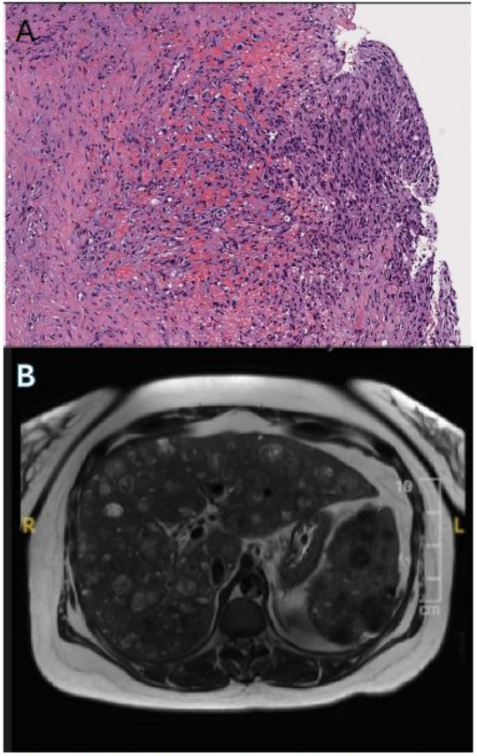
Figure: Figure A: Photomicrograph of liver showing malignant endothelial proliferation with irregular, blood-filled vascular spaces and areas of hemorrhage and necrosis, typical of high-grade hepatic spindle cell angiosarcoma. Figure B: axial MRI of the abdomen, revealing multiple liver masses, with the most significant right hepatic lobe lesion measuring approximately 8.0 x 9.2 cm.
Disclosures: Ahmad Afzal indicated no relevant financial relationships. Saad Nadeem indicated no relevant financial relationships. Abdul Basit Afzal indicated no relevant financial relationships. Apaar Dadlani indicated no relevant financial relationships. Scott Berger indicated no relevant financial relationships. Donghwa Baek indicated no relevant financial relationships. Sudha Kodali: ASTRAZENECA – Advisory Committee/Board Member. GILEAD – Advisor or Review Panel Member, Advisory Committee/Board Member, Speakers Bureau. SIRTEX – Advisor or Review Panel Member.
Ahmad Afzal, MD1, Saad Nadeem, MD1, Abdul Basit Afzal, MBBS2, Apaar Dadlani, MD1, Scott Berger, MD1, Donghwa Baek, MD1, Sudha Kodali, MD1. P6028 - A Rare and Diagnostically Challenging Case of Spindle Cell Liver Angiosarcoma, ACG 2025 Annual Scientific Meeting Abstracts. Phoenix, AZ: American College of Gastroenterology.