Madison Piao, 1, Christopher N. Ton, 2, Ashwini Mulgaonkar, MD3 1University of California Berkeley, San Diego, CA; 2Kaiser Permanente, Vacaville, CA; 3Kaiser Permanente, Sacramento, CA Introduction: Small bowel bleeding is uncommon and accounts for 5-10% of cases of GI bleeding. In this clinical case, we describe a case of small bowel bleeding that was identified through a collaborative effort between Gastroenterology and Surgery.
Case Description/
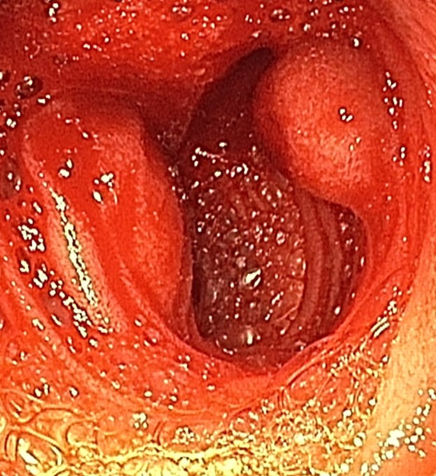
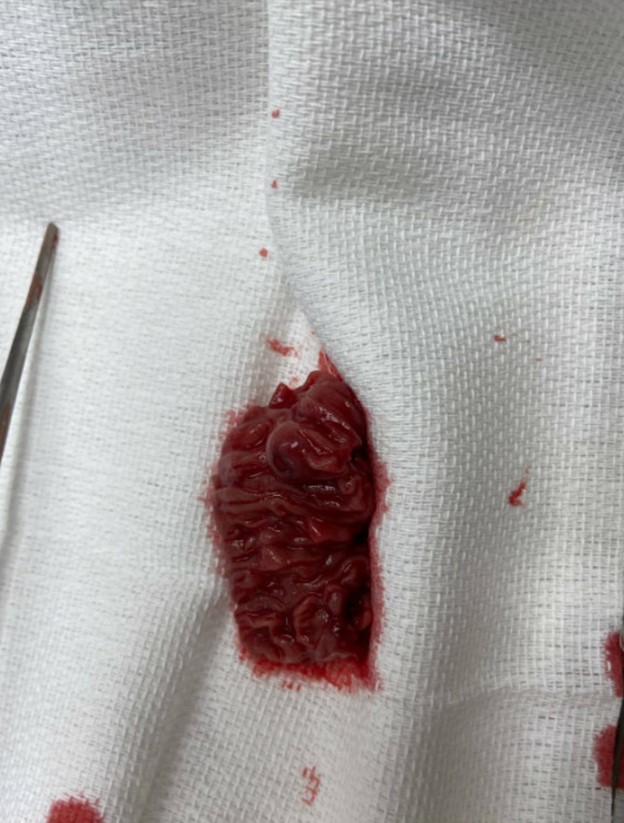
Methods: A 22-year-old man with a history of obesity with a BMI of 37 presented to the Emergency Room with presyncope and multiple weeks of diarrhea with dark stools that worsened 4 days before presentation. Initial labs showed a drop in hemoglobin (Hgb) from baseline of 12.8 g/dL to 6.1g/dL. After resuscitation, he underwent upper endoscopy that was normal. He subsequently developed large volume hematochezia with associated hemodynamic instability. Urgent CT angiogram of the abdomen showed active extravasation at the proximal jejunum. Patient underwent a laparotomy with intra- operative enteroscopy. Our surgeon(s) assisted in the push enteroscopy with exploratory laparotomy by providing manual pressure to the intestinal wall and colonoscope, allowing the advancement of the scope to the ileum. A small mucosal mass was identified by palpation in the proximal jejunum by the surgeons. The colonoscope was advanced back to this area, where a visible vessel was seen and actively bleeding from an ulcerated portion of a submucosal mass. The lesion was transected, and a functional end-to-end anastomosis was performed. The surgical pathology showed a subepithelial organizing hematoma with ulceration. The patient had an uncomplicated post-operative course without any episodes of additional GI bleeding. Discussion: Non-traumatic hematomas in the small bowel are exceedingly rare, with variable presentation, but they typically resolve with conservative management(1). They are more commonly seen in elderly patients on anticoagulation. The underlying cause of the intramural hematoma is often unclear, as it was in this case, with the differential including arteriovenous malformation, Dieulafoy, or spontaneous hematoma. Here, we discuss a rare case of non-traumatic small bowel hematoma as a cause of large volume intraluminal bleeding that required endoscopic evaluation with surgical resection for definitive treatment in a young patient without any coagulopathies.
References: 1. Abbas MA, Collins JM, Olden KW, Kelly KA. Spontaneous Intramural Small-Bowel Hematoma: Clinical Presentation and Long-term Outcome. Arch Surg. 2002;137(3):306–310. doi:10.1001/archsurg.137.3.306
Figure: Endoscopic image of an ulcerated hematoma in the proximal jejunum with active bleeding.
Figure: Gross surgical specimen of the resected jejunum.
Disclosures: Madison Piao indicated no relevant financial relationships. Christopher Ton indicated no relevant financial relationships. Ashwini Mulgaonkar indicated no relevant financial relationships.
Madison Piao, 1, Christopher N. Ton, 2, Ashwini Mulgaonkar, MD3. P6255 - An Unusual Case of Gastrointestinal Bleeding, ACG 2025 Annual Scientific Meeting Abstracts. Phoenix, AZ: American College of Gastroenterology.