Alex Lee, MD AdventHealth, Orlando, FL Introduction: Ascites is a common complication of cirrhosis due to portal hypertension and hepatic insufficiency. In rare cases, ascitic fluid can migrate into a coexisting inguinal hernia and accumulate in the scrotum, leading to significant enlargement. Scrotal drainage is infrequently performed, as most reported cases involve hydroceles rather than ascitic fluid accumulation within the scrotum secondary to an inguinal hernia, as seen in our case.
Case Description/
Methods: A 67-year-old man with cirrhosis (Child-Pugh Class B, MELD 10) and a longstanding inguinal hernia presented with progressive scrotal swelling, pain, and skin breakdown. Physical exam revealed a massively enlarged, erythematous scrotum with buried penis and visible wound drainage. Imaging showed a large fluid-filled scrotal sac extending from the inguinal hernia with no detectable abdominal ascites. Lab were significant for elevated WBC and Hepatitis C infection with a viral load of 1210375 IU/mL. Other labs including ALT, alkaline phosphatase, bilirubin, PT/PTT, glucose, BUN, AFP, and a urine drug screen were within normal limits. Hepatitis serologies were also positive for anti-HBc and IgG anti-HAV, indicating past infections with Hepatitis B and Hepatitis A, respectively. Wound cultures grew Pantoea agglomerans. Initial management included diuretics (furosemide, spironolactone), topical antibiotics, and use of a PureWick catheter due to inability to place a Foley. Ultrasound-guided scrotal centesis was performed using a pigtail catheter and vacuum suction, removing 9.4 liters of clear yellow ascitic fluid. Fluid analysis was consistent with transudative ascites without signs of active spontaneous bacterial peritonitis (albumin 0.8, nucleated cells 49). Post-procedure, scrotal size was reduced, and the patient was discharged with continued diuretics and plans for Hepatitis C treatment and eventual hernia repair. Discussion: This case illustrates a rare presentation of scrotal ascites without intra-abdominal fluid, requiring multidisciplinary management. The patient’s surgical risk was evaluated using the VOCAL-Penn score (open surgery: 90-day mortality 16.3%). Wound care involved advanced dressing techniques, and potential TIPS is being considered to prevent recurrence. Only few cases have reported scrotal centesis for ascites, with significantly less volume drained. This case highlights the importance of early recognition, fluid control, and coordinated long-term care in cirrhotic patients with hernias and unusual fluid distribution.
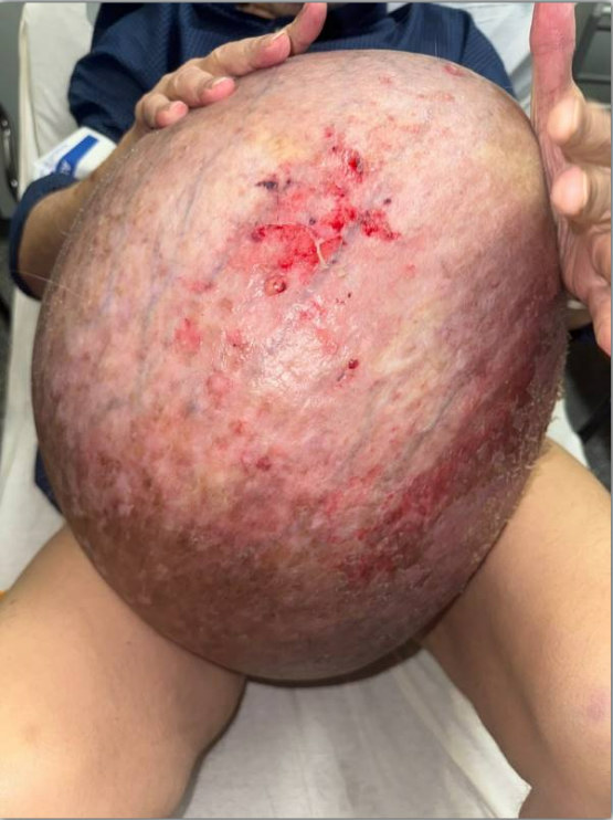
Figure: On initial presentation, the scrotum was significantly enlarged. The scrotal skin appeared maroon, tender, and exhibited nonblanchable erythema, consistent with friction-related skin breakdown. Multiple wounds and areas of redness with some secretion were noted. The penis and urethral meatus were not visible due to burial within the massively enlarged scrotum.
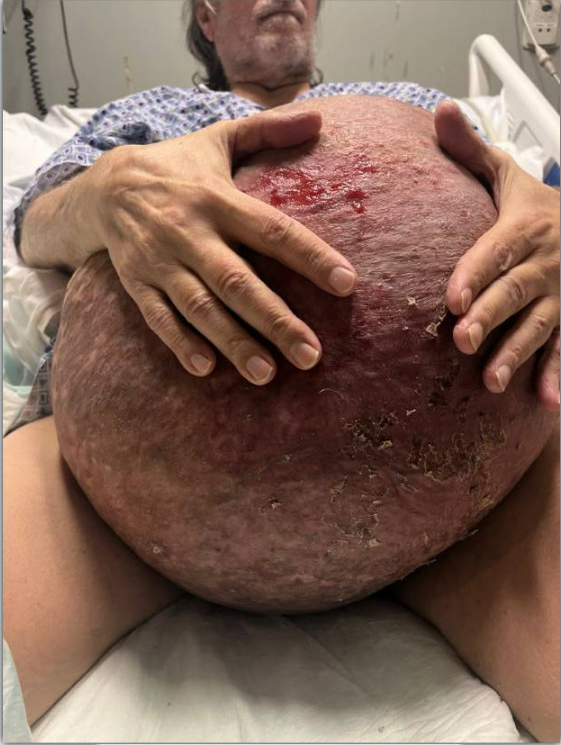
Figure: After removal of 9.4 liters of clear yellow ascitic fluid. The scrotum remained severely enlarged, but the size and skin tension were significantly reduced.
Disclosures: Alex Lee indicated no relevant financial relationships.
Alex Lee, MD. P6238 - Inguinal Hernia With Massive Fluid Collection in the Scrotum in a Cirrhotic Patient, ACG 2025 Annual Scientific Meeting Abstracts. Phoenix, AZ: American College of Gastroenterology.