HCA Healthcare Riverside Community Hospital Riverside, CA
Regis Lee, DO1, Raj Gulati, DO2, Nishani Wickramarachchi, DO1, Fadi Totah, DO2, Alex Prevallet, DO1, Scott Lee, MD2 1HCA Healthcare Riverside Community Hospital, Riverside, CA; 2Riverside Community Hospital, Riverside, CA Introduction: Malignancy of the small intestine accounts for a rare subsection of gastrointestinal carcinoma encompassing up to 3% of gastrointestinal cancer patients, with terminal ileum adenocarcinoma appearing at the lowest rate. These cancers are poorly diagnosed given their lack of specificity in the clinical setting. This case presents a patient with a history of recurrent small bowel obstruction (SBO) who was found to have adenocarcinoma of the small intestine.
Case Description/
Methods: A 70-year-old female with a history of hypertension, hyperlipidemia, and multiple small bowel obstructions (SBO) presented with one month of abdominal pain and concern for recurrent SBO. At the outside hospital she had a CT abdomen and pelvis showing mildly dilated loops of small bowel in the mid abdomen measuring up to 3.6 cm with areas of small bowel wall thickening representing possible small bowel obstruction and soft tissue nodularity in the LUQ representing possible metastatic omental disease. Her last colonoscopy was over 10 years ago. She denied any family history of inflammatory bowel disease or alarming features. Colonoscopy was performed for concerns of inflammatory bowel disease revealing a 20-25 mm submucosal appearing lesion at the base of the cecum and a normal appearing mucosa in the terminal ileum that appeared externally compressed. Biopsies from the cecal lesion were negative for malignancy, however a biopsy from the terminal ileum revealed goblet cells suspicious of adenocarcinoma of the appendix. She was then evaluated by surgery however she was deemed not a surgical candidate. She was then discharged in stable condition and instructed to follow up with oncology for initiation of chemotherapy. Discussion: Adenocarcinoma of the terminal ileum and appendix are exceedingly rare, often presenting with vague gastrointestinal symptoms, which can delay diagnosis and treatment. In this case, the patient's recurrent episodes of SBO and nonspecific imaging findings initially raised suspicion for Crohn’s disease—a common diagnostic pitfall in terminal ileal cancers. This case highlights the workup of small bowel malignancies and how early recognition can improve outcomes.
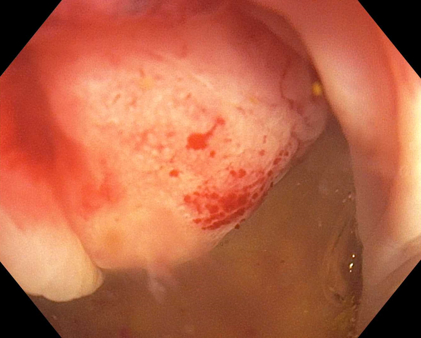
Figure: 20 to 25 mm submucosal lesion present at base of cecum.
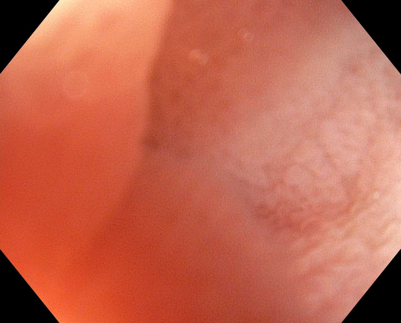
Figure: Terminal ileum unable to distend despite air insufflation and water. Questionable extrinsic compression from cecal lesion.
Disclosures: Regis Lee indicated no relevant financial relationships. Raj Gulati indicated no relevant financial relationships. Nishani Wickramarachchi indicated no relevant financial relationships. Fadi Totah indicated no relevant financial relationships. Alex Prevallet indicated no relevant financial relationships. Scott Lee indicated no relevant financial relationships.
Regis Lee, DO1, Raj Gulati, DO2, Nishani Wickramarachchi, DO1, Fadi Totah, DO2, Alex Prevallet, DO1, Scott Lee, MD2. P6276 - Unmasking a Rare Malignancy: Terminal Ileum Adenocarcinoma Diagnosed After Recurrent Small Bowel Obstruction, ACG 2025 Annual Scientific Meeting Abstracts. Phoenix, AZ: American College of Gastroenterology.