Ivana Rubenstein, DO1, Gautam Anand, MD1, Satya Singh, MD1, Aryama Sharma, MD2 1Broward Health Medical Center, Fort Lauderdale, FL; 2Broward Health North, Deerfield Beach, FL Introduction: Jejunal diverticulosis, a rare condition involving diverticula in the jejunum, is often found incidentally or when complicated by diverticulitis. While more common in the colon, jejunal diverticulosis poses diagnostic challenges due to its rarity and nonspecific symptoms. Imaging, particularly CT, is crucial for diagnosing jejunal diverticulitis and guiding management.
Case Description/
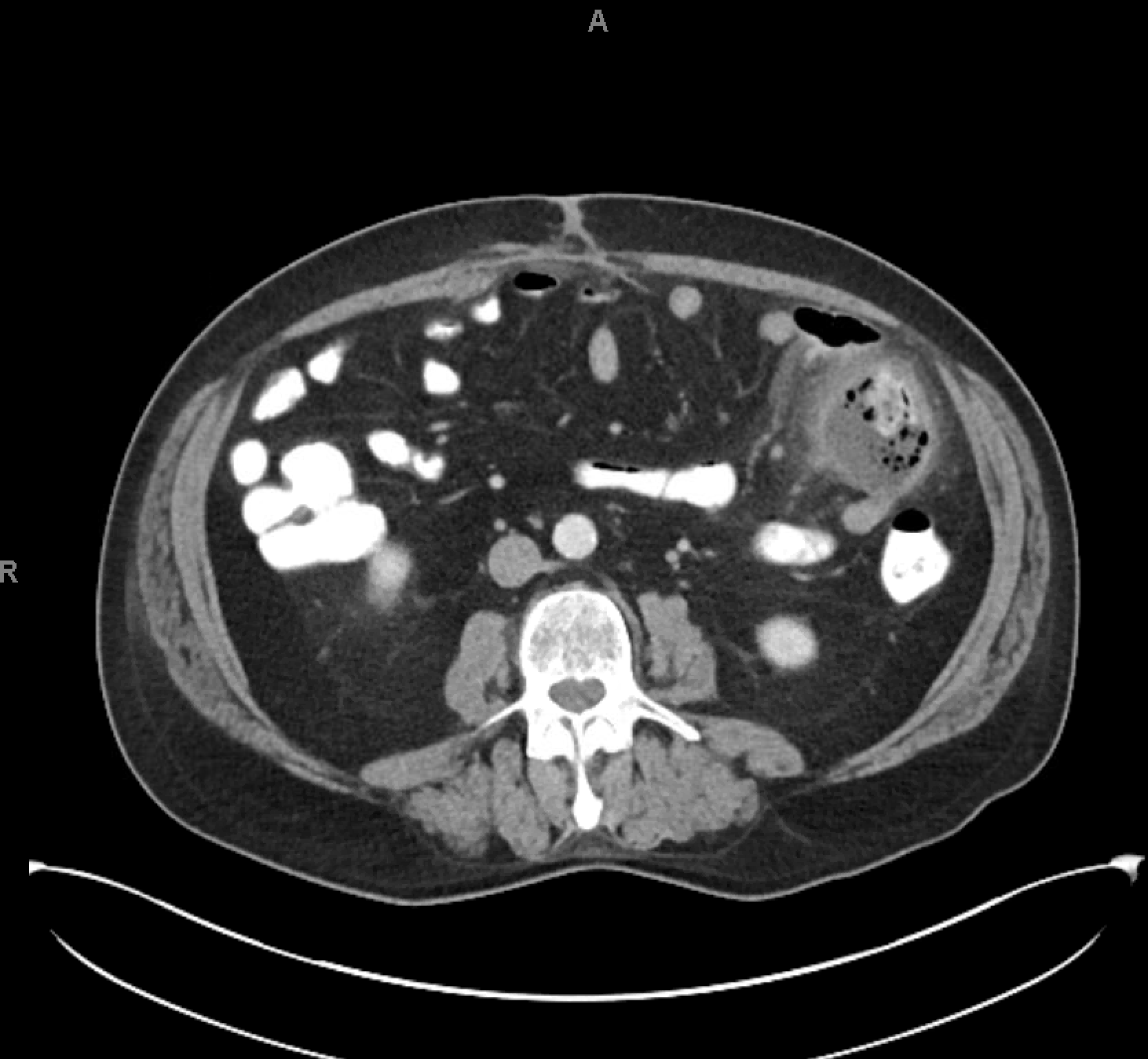
Methods: A 70-year-old male with history of hypertension developed acute left lower abdominal pain while on vacationing in Jamaica. The patient opted to return to the U.S. for care, where CT with IV contrast confirmed multiple small jejunal diverticula and a 4.5 x 4.1 cm circular mass with fluid and debris. The mass communicated with the bowel, suggesting diverticulitis or, less likely, a contained perforation with abscess.
Follow-up CT with oral contrast showed a 5 cm x 5 cm rim-enhancing structure near the jejunum containing succus, oral contrast, and air, with surrounding fat induration and adjacent small lymph nodes. These findings were consistent with jejunal diverticulitis complicated by an abscess. No free air or obstruction was present. The patient underwent robotic-assisted laparoscopic surgery, including lysis of adhesions, partial enterectomy with primary anastomosis, and partial omentectomy. He received an IV antibiotic course, clinically improved, and was discharged after recovery. Discussion: Jejunal diverticulosis, while uncommon, can range from asymptomatic to presenting with acute abdominal pain, nausea, vomiting, and gastrointestinal bleeding. Complications such as diverticulitis, perforation, or abscess formation can occur, requiring prompt diagnosis and intervention. CT imaging plays a pivotal role in diagnosing jejunal diverticulitis by revealing diverticula, associated inflammatory changes, and potential for abscess formation. In this case, CT with oral and IV contrast was essential in differentiating jejunal diverticulitis from other abdominal pathologies, such as small bowel perforation or mesenteric ischemia.
Jejunal diverticulitis is typically managed conservatively with antibiotics in uncomplicated cases. However, surgical intervention is required when abscess or perforation is present. Surgical options include bowel resection, primary anastomosis, and abscess drainage, depending on the severity. Our patient's successful recovery following robotic-assisted laparoscopic surgery highlights the effectiveness of minimally invasive techniques in managing complicated jejunal diverticulitis.
Figure: Figure 1: CT image with IV and oral contrast of abdomen showing multiple small jejunal diverticula and a 5 cm x 5 cm rim-enhancing mass near the jejunum. These findings are consistent with jejunal diverticulitis complicated by an abscess.
Disclosures: Ivana Rubenstein indicated no relevant financial relationships. Gautam Anand indicated no relevant financial relationships. Satya Singh indicated no relevant financial relationships. Aryama Sharma indicated no relevant financial relationships.
Ivana Rubenstein, DO1, Gautam Anand, MD1, Satya Singh, MD1, Aryama Sharma, MD2. P6274 - Acute Jejunal Diverticulitis Complicated by Abscess: A Diagnostic and Management Challenge, ACG 2025 Annual Scientific Meeting Abstracts. Phoenix, AZ: American College of Gastroenterology.

 photo")