University of Illinois College of Medicine Peoria, IL
Navjit Singh, MD, Ankita Nekkanti, MD, Imran Balouch, MD University of Illinois College of Medicine, Peoria, IL Introduction: Hepatocellular adenoma (HCA) is a benign liver tumor that is a rare entity in males. When present, they are associated with malignant transformation of ~4.2%. HCA is predominantly found in females, with higher incidence in those using oral contraceptives. ~10% of all HCAs occur in men and the incidence has been rising over the past few decades. HCA-related risk factors include anabolic steroid use, obesity, and metabolic syndrome. In males with HCA, surgical resection is generally accepted, irrespective of size, based on the high risk and difficult differentiation with hepatocellular carcinoma (HCC).
Case Description/
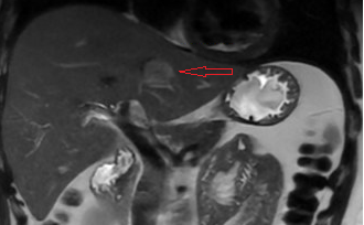
Methods: A 17-year-old Hispanic male with obesity was referred for asymptomatic mildly elevated AST (52 U/L). Abdominal ultrasound revealed a 2.8 × 2.7 cm hypoechoic left liver lesion, along with features of fatty liver. He denied alcohol use, and had no family history of liver disease. MRI confirmed a 2.2 × 2.6 cm segment II lesion with arterial enhancement and delayed washout, suspicious for adenoma or carcinoma. Alpha-fetoprotein (AFP) was normal. CT-guided biopsy confirmed an inflammatory HCA. At a liver tumor conference, resection was recommended due to his sex, but surveillance was chosen until the lesion is appropriate for surgery. Discussion: HCA is a rare benign liver tumor, typically seen in women of reproductive age and linked to estrogen exposure. Its occurrence in males, especially teenagers, is uncommon and carries a higher risk of malignant transformation. HCAs are often asymptomatic and found on imaging, which, along with biopsy, is essential for diagnosis. This young patient’s lesion was classified as an inflammatory HCA, accounting for ~30% of cases. In males, up to 47% of HCAs may transform into hepatocellular carcinoma, prompting surgical resection regardless of size. In contrast, female patients are often managed based on subtype or lesion size ( >5 cm). Here, due to the lesion’s small size, a multidisciplinary team opted for surveillance, with planned resection when appropriate. Normal AFP and absence of risk factors supported this approach. This case highlights the need for early detection, accurate classification, and tailored management of HCA in males.
Figure: Figure 1: MRI showing segment II lesion measuring 2.2 x 2.6 cm (arrow) with suspicious characteristics of adenoma, including early arterial enhancement and delayed phase washout.
Disclosures: Navjit Singh indicated no relevant financial relationships. Ankita Nekkanti indicated no relevant financial relationships. Imran Balouch indicated no relevant financial relationships.
Navjit Singh, MD, Ankita Nekkanti, MD, Imran Balouch, MD. P6125 - Hepatocellular Adenoma in a Teenage Male, ACG 2025 Annual Scientific Meeting Abstracts. Phoenix, AZ: American College of Gastroenterology.