Salman Ahmed, DO1, Cristina Angelo, MD1, Adam Peyton, DO1, Jillian Grau, MD2 1Lehigh Valley Health Network, Allentown, PA; 2LVHN, Allentown, PA Introduction: Leiomyomas are benign, smooth muscle neoplasms which often arise in the genitourinary (GU) and gastrointestinal (GI) systems. Leiomyomas infrequently spread to distal sites; a phenomenon known as Benign Metastasizing Leiomyomas (BML). To our knowledge, there’s only one other reported case of BML which presented with isolated hepatic involvement. Here, we report a case of BML with liver involvement identified twelve years after hysterectomy for uterine fibroids.
Case Description/
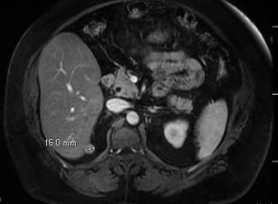
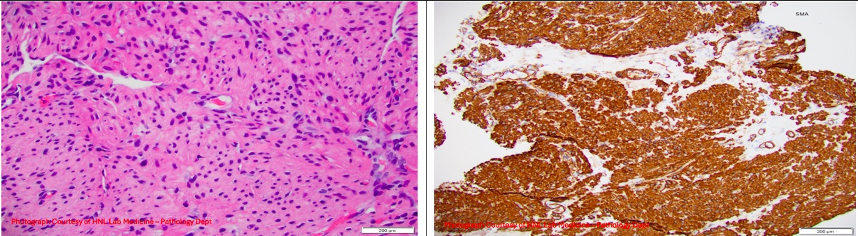
Methods: A 61-year-old female with a history of ductal carcinoma in situ of the right breast status post lumpectomy and radiation, uterine fibroids status post hysterectomy, hypertension, and hepatic steatosis presented for evaluation of a liver lesion and one year of nonspecific right upper abdominal pain. An MRI lumbar spine revealed an incidental finding of a 1.6 cm T2 hyperintense lesion adjacent to the liver. Prior abdominal imaging only revealed hepatic steatosis. Liver function tests noted total bilirubin 0.96, AST 26, ALT 32, alkaline phosphatase 53. MRI abdomen confirmed a 16 mm exophytic liver lesion (Figure 1). A biopsy was performed, and pathology was consistent with benign smooth muscle tissue. The patient underwent a repeat biopsy for confirmatory testing, and pathology identified bland appearing spindle cell lesion without atypia, necrosis, and mitotic activity findings compatible with a leiomyoma (Figure 2). Immunostaining was positive for desmin, SMA, and h-caldesmon which supported the diagnosis. Given the benign histological finding and stable size, the decision was made to monitor the lesion for growth with imaging. Discussion: Primary and metastasizing liver leiomyomas are exceedingly rare. Of the cases described most incidents of BML discuss lung involvement, and reported cases on hepatic BML is limited. There are no standard treatment guidelines for BML, but they generally carry a favorable prognosis and can be monitored for growth. In some cases, estrogen modulators, progestins, and VEGF inhibitors have been used for tumor regression. This case emphasizes the importance of considering hepatic BML in the differential of benign liver lesions and the importance of immunohistochemistry to help differentiate benign lesions from malignancy.
The use of generative AI such as ChatGPT was limited to assist with grammar and clarity during the early drafting stage. The final content including all clinical interpretation and language was authored and reviewed independently.
Figure: 16 mm exophytic solid lesion arising from the medial aspect of hepatic segment 6.
Disclosures: Salman Ahmed indicated no relevant financial relationships. Cristina Angelo indicated no relevant financial relationships. Adam Peyton indicated no relevant financial relationships. Jillian Grau indicated no relevant financial relationships.
Salman Ahmed, DO1, Cristina Angelo, MD1, Adam Peyton, DO1, Jillian Grau, MD2. P5995 - Latent Leiomyoma: A Delayed Diagnosis of Metastasizing Leiomyoma, ACG 2025 Annual Scientific Meeting Abstracts. Phoenix, AZ: American College of Gastroenterology.