Corewell Health William Beaumont University Hospital Royal Oak, MI
Marko Kozyk, MD1, Rabin Neupane, MD2, Kateryna Strubchevska, MD1, Laith H. Jamil, MD, FACG3 1Corewell Health William Beaumont University Hospital, Royal Oak, MI; 2AnMed Gastroenterology, Greenville, SC; 3Oakland University William Beaumont School of Medicine, Rochester, MI Introduction: One rare complication of percutaneous endoscopic transgastric jejunostomy tube (PEG-J) placement is a transcolonic misplacement of the tube. We present a technique of endoscopic closure of gastrocolic (GC) and colocutaneous (CC) fistulas, which are caused by the misplacement of the PEG-J tube.
Case Description/
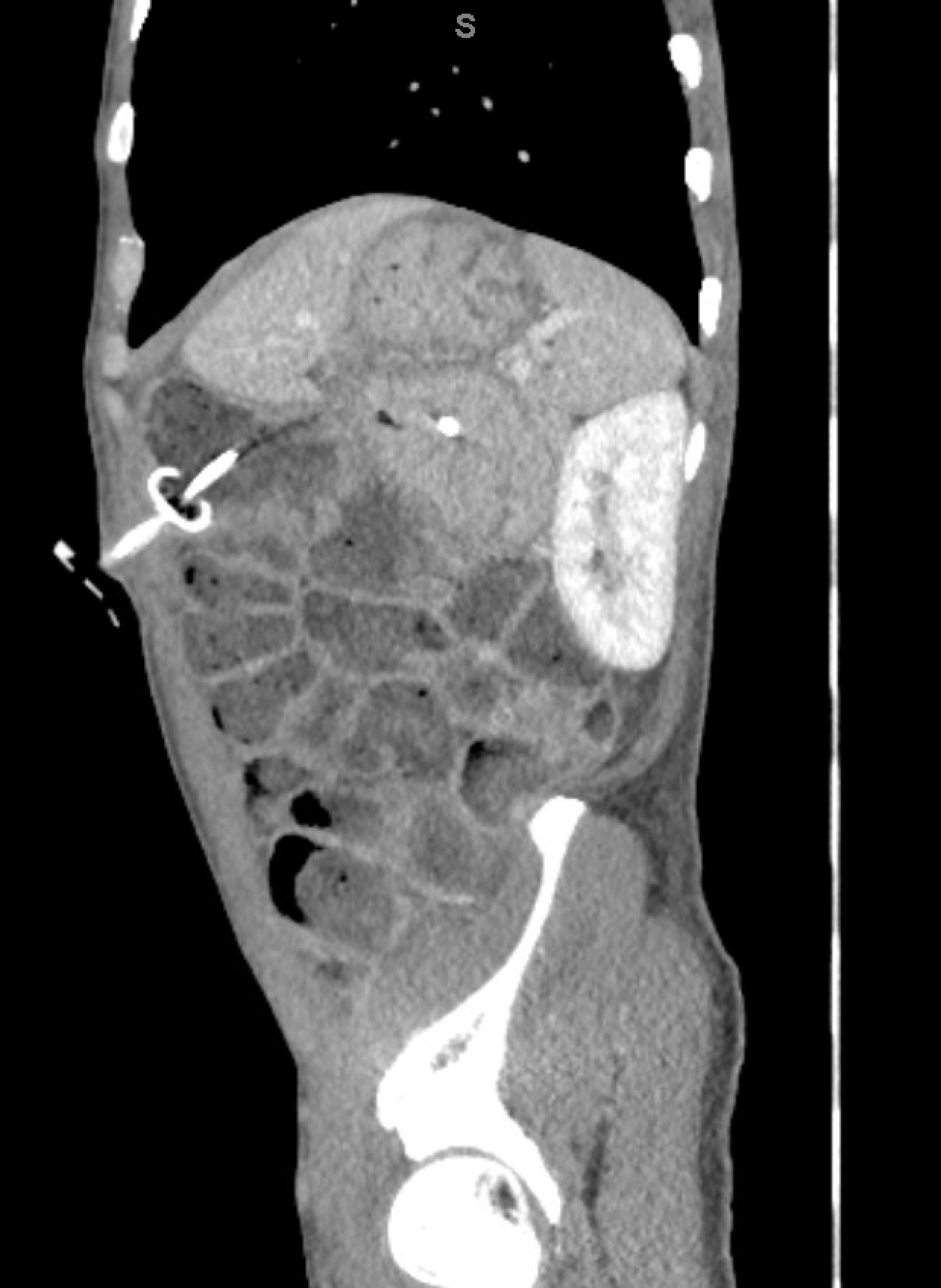
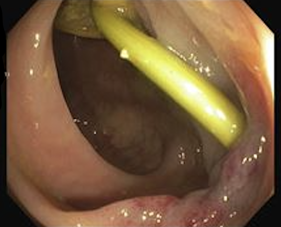
Methods: A 49-year-old male with a past medical history significant for duodenal volvulus status post exploratory laparotomy with duodenojejunostomy and a recent EGD with PEG-J placement presented with complaints of abdominal pain. The proximal portion of the PEG-J tube appeared to be within the loops of the colon in the left upper quadrant, subsequently traversing into the stomach and terminating at the duodenum (Figure 1). On EGD, the PEG- J tube was found coming through the gastric body. The bumper was not found in the stomach, but the J portion of the tube was extending to the duodenum. A hemostatic clip was placed right above the tract in the gastric body to mark the location. This was followed by a colonoscopy. The PEG-J tube was seen in the transverse colon traversing the colonic lumen (Figure 2). The bumper was located within the lumen and the J portion was seen extending through the colonic mucosa to the stomach. The scope was then withdrawn, and an over-the-scope clip (OTSC) was placed and reinserted. First, an alligator rat-toothed forceps was used to grasp the edge where the J tube exited the colon wall into the stomach. The J portion of the PEG J tube was then removed from the outside. Once removed, the defect grasped by the alligator forceps was pulled into the OTSC which was then deployed. For the removal of the G portion of the PEG J tube, a straight Jagwire was passed through the PEG tube from the external opening. The PEG tube was then pushed into the colon from outside with the wire in place. The edge of the defect was grasped with an alligator rat-toothed forceps and pulled into the OTSC. Once the position was confirmed with fluoroscopy, the wire was pulled out and the colo-cutaneous fistula was closed with OTSC. The PEG tube then was taken out with forceps. This was followed by an EGD where the fistula was noted just below the endo clip which was placed initially for marking. The defect was then closed with five endoclips. Discussion: Our case demonstrates that endoscopic technique can safely close gastrocolic and colocutaneous fistulas using clips in a patient, who does not have any signs of overt intraabdominal infection and is not a high-risk surgical candidate.
Figure: CT abdomen pelvis (sagittal view).
Figure: Colonoscopy findings of gastrojejunal tube.
Disclosures: Marko Kozyk indicated no relevant financial relationships. Rabin Neupane indicated no relevant financial relationships. Kateryna Strubchevska indicated no relevant financial relationships. Laith Jamil indicated no relevant financial relationships.
Marko Kozyk, MD1, Rabin Neupane, MD2, Kateryna Strubchevska, MD1, Laith H. Jamil, MD, FACG3. P3564 - Endoscopic Management of Misplaced Percutaneous Endoscopic Gastrojejunal Tube, ACG 2025 Annual Scientific Meeting Abstracts. Phoenix, AZ: American College of Gastroenterology.

 photo")