Monday Poster Session
Category: GI Bleeding
P3136 - An Elusive Small Bowel GIST in an Atypical Patient
Monday, October 27, 2025
10:30 AM - 4:00 PM PDT
Location: Exhibit Hall

Mrudula Bandaru, MD (she/her/hers)
Department of Medicine, George Washington University School of Medicine and Health Sciences
Washington, DC
Presenting Author(s)
Mrudula Bandaru, MD1, Romy Chamoun, MD2, Kathryn Hobbs, MD3, Khashayar Vaziri, MD2, Marie L. Borum, MD, EdD, MPH, FACG4, Samuel A. Schueler, MD4
1Department of Medicine, George Washington University School of Medicine and Health Sciences, Washington, DC; 2George Washington University School of Medicine and Health Sciences, Washington, DC; 3Inova Fairfax Medical Campus, Fairfax, VA; 4Division of Gastroenterology and Liver Disease, Department of Medicine, George Washington University School of Medicine and Health Sciences, Washington, DC
Introduction: Gastrointestinal stromal tumors (GISTs) are rare mesenchymal tumors that may arise throughout and around the gastrointestinal (GI) tract with median age of diagnosis between 65-69 years. We present a case of an otherwise healthy 43-year-old male presenting with intermittent melena, eventually found to have a jejunal GIST.
Case Description/
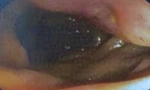
Methods: A healthy 43-year-old male presented with intermittent melena and iron deficiency anemia. Upper endoscopy and colonoscopy were unrevealing. He presented again with melena at which time push enteroscopy was also unrevealing. A capsule endoscopy revealed active proximal small bowel bleeding approximated to the distal duodenum, thus a repeat push enteroscopy was conducted. This revealed a proximal jejunal ulceration that was just out of reach of the endoscope (Figure 1). A hemostatic clip was placed near the lesion. Finally, a double-balloon enteroscopy identified a 2.5 – 3.0cm ulcerated jejunal mass. Biopsy revealed bland spindle cells. Immunostaining revealed positive DOG1 and CD117 and negative desmin stain, all consistent with a GIST. CT abdomen/pelvis did not reveal metastatic disease. In a subsequent encounter, he again developed melena requiring admission, red blood cell transfusions and urgently underwent a laparoscopic small bowel resection of a 3.0 cm grade 1 GIST. He is doing well at follow-up without anemia or further episodes of melena.
Discussion: GISTs account for 0.04% of all GI tumors and typically present in older adults. GISTs are found in the stomach (60%), small intestine (30%), and rarely in the esophagus, colon, rectum, or outside of the GI tract. Metastatic disease is present at diagnosis in 10-20% of cases. They may lead to significant GI bleeding. Our patient suffered through multiple episodes of melena requiring multiple admissions and blood transfusions over the course of weeks-to-months while the above diagnostic evaluation took place. This case highlights the importance of recognizing that GISTs may occur in younger adults or even children and may cause life-threatening GI bleeding. A higher index of suspicion and more aggressive initial diagnostic approach may have resulted in decreased morbidity prior to ultimate curative resection.

Figure: Figure 1: Ulcer in the proximal jejunum identified during push enteroscopy
Disclosures:
Mrudula Bandaru indicated no relevant financial relationships.
Romy Chamoun indicated no relevant financial relationships.
Kathryn Hobbs indicated no relevant financial relationships.
Khashayar Vaziri indicated no relevant financial relationships.
Marie Borum indicated no relevant financial relationships.
Samuel Schueler indicated no relevant financial relationships.
Mrudula Bandaru, MD1, Romy Chamoun, MD2, Kathryn Hobbs, MD3, Khashayar Vaziri, MD2, Marie L. Borum, MD, EdD, MPH, FACG4, Samuel A. Schueler, MD4. P3136 - An Elusive Small Bowel GIST in an Atypical Patient, ACG 2025 Annual Scientific Meeting Abstracts. Phoenix, AZ: American College of Gastroenterology.
1Department of Medicine, George Washington University School of Medicine and Health Sciences, Washington, DC; 2George Washington University School of Medicine and Health Sciences, Washington, DC; 3Inova Fairfax Medical Campus, Fairfax, VA; 4Division of Gastroenterology and Liver Disease, Department of Medicine, George Washington University School of Medicine and Health Sciences, Washington, DC
Introduction: Gastrointestinal stromal tumors (GISTs) are rare mesenchymal tumors that may arise throughout and around the gastrointestinal (GI) tract with median age of diagnosis between 65-69 years. We present a case of an otherwise healthy 43-year-old male presenting with intermittent melena, eventually found to have a jejunal GIST.
Case Description/
Methods: A healthy 43-year-old male presented with intermittent melena and iron deficiency anemia. Upper endoscopy and colonoscopy were unrevealing. He presented again with melena at which time push enteroscopy was also unrevealing. A capsule endoscopy revealed active proximal small bowel bleeding approximated to the distal duodenum, thus a repeat push enteroscopy was conducted. This revealed a proximal jejunal ulceration that was just out of reach of the endoscope (Figure 1). A hemostatic clip was placed near the lesion. Finally, a double-balloon enteroscopy identified a 2.5 – 3.0cm ulcerated jejunal mass. Biopsy revealed bland spindle cells. Immunostaining revealed positive DOG1 and CD117 and negative desmin stain, all consistent with a GIST. CT abdomen/pelvis did not reveal metastatic disease. In a subsequent encounter, he again developed melena requiring admission, red blood cell transfusions and urgently underwent a laparoscopic small bowel resection of a 3.0 cm grade 1 GIST. He is doing well at follow-up without anemia or further episodes of melena.
Discussion: GISTs account for 0.04% of all GI tumors and typically present in older adults. GISTs are found in the stomach (60%), small intestine (30%), and rarely in the esophagus, colon, rectum, or outside of the GI tract. Metastatic disease is present at diagnosis in 10-20% of cases. They may lead to significant GI bleeding. Our patient suffered through multiple episodes of melena requiring multiple admissions and blood transfusions over the course of weeks-to-months while the above diagnostic evaluation took place. This case highlights the importance of recognizing that GISTs may occur in younger adults or even children and may cause life-threatening GI bleeding. A higher index of suspicion and more aggressive initial diagnostic approach may have resulted in decreased morbidity prior to ultimate curative resection.

Figure: Figure 1: Ulcer in the proximal jejunum identified during push enteroscopy
Disclosures:
Mrudula Bandaru indicated no relevant financial relationships.
Romy Chamoun indicated no relevant financial relationships.
Kathryn Hobbs indicated no relevant financial relationships.
Khashayar Vaziri indicated no relevant financial relationships.
Marie Borum indicated no relevant financial relationships.
Samuel Schueler indicated no relevant financial relationships.
Mrudula Bandaru, MD1, Romy Chamoun, MD2, Kathryn Hobbs, MD3, Khashayar Vaziri, MD2, Marie L. Borum, MD, EdD, MPH, FACG4, Samuel A. Schueler, MD4. P3136 - An Elusive Small Bowel GIST in an Atypical Patient, ACG 2025 Annual Scientific Meeting Abstracts. Phoenix, AZ: American College of Gastroenterology.
