Madeline Washburn-Rodriguez, DO, My Nguyen, DO, Luke Kennedy, MD, James T. Sing, DO, Niket Sonpal, MD Baylor Scott & White Medical Center, Temple, TX Introduction: Gastric adenomas comprise less than 10% of all gastric polyps, and pyloric gland adenomas (PGAs) are estimated to account for only 2.7%. PGAs are typically described as flat or polypoid, and the average size is estimated to be approximately 1.7-cm. Though current literature mentions the possibility of larger and/or ulcerated PGAs, the incidence remains unclear. Here, we present a unique occurrence of a large, ulcerated gastric mass initially concerning for malignancy with pathology consistent with a benign pyloric gland adenoma.
Case Description/
Methods: Our patient was a 63-year-old female who presented after two days of melena and hematemesis. She had no prior history of gastrointestinal bleeds; however, one month prior to this encounter, she was admitted for new-onset atrial fibrillation for which anticoagulation was initiated.
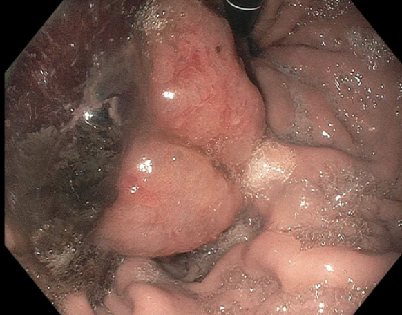
Initial workup was significant for a hemoglobin of 6.4 for which she was transfused. Gastroenterology was consulted. An upper endoscopy discovered an ulcerated, 4-cm pedunculated gastric mass with an adherent clot (Figure 1). There was no active bleeding, however old blood was visualized in the stomach. Biopsies were obtained though resection was delayed due to recent anticoagulation.
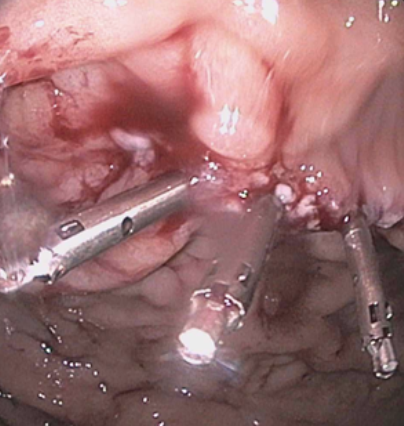
Repeat upper endoscopy was performed for removal of the large gastric polyp which was removed by electrocautery in piecemeal fashion (Figure 2). The pathology reports of the biopsy and resection showed fragments of a pyloric gland adenoma without signs of high-grade dysplasia. Discussion: Pyloric gland adenomas are rare, accounting for less than 3% of all gastric polyps. They are typically small and found incidentally. Our patient's very large, ulcerated lesion found during upper endoscopy was highly concerning for malignancy; however, the pathology report showed no sign of high-grade dysplasia. The patient likely had the adenoma for many years, and ulceration with bleeding was likely a result of recent initiation of anticoagulation.
Pyloric gland adenomas are benign, precancerous lesions; however, the rate of transformation into adenocarcinoma ranges from 12.2% to nearly 50%. Given the risk of transformation, the American Society of Gastrointestinal Endoscopy recommends repeat endoscopy one year post resection and surveillance endoscopy every 3-5 years thereafter. Our patient is scheduled in our clinic to discuss follow-up procedures. Acute blood loss anemia secondary to a pyloric gland adenoma is a unique presentation of an otherwise benign gastric mass.
Figure: Retroflexed view of the pedunculated PGA with an adherent clot.
Figure: Status post piecemeal resection and closure with 3 hemoclips.
Disclosures: Madeline Washburn-Rodriguez indicated no relevant financial relationships. My Nguyen indicated no relevant financial relationships. Luke Kennedy indicated no relevant financial relationships. James Sing indicated no relevant financial relationships. Niket Sonpal indicated no relevant financial relationships.
Madeline Washburn-Rodriguez, DO, My Nguyen, DO, Luke Kennedy, MD, James T. Sing, DO, Niket Sonpal, MD. P3018 - The Not so Benign-Appearing Pyloric Gland Adenoma, ACG 2025 Annual Scientific Meeting Abstracts. Phoenix, AZ: American College of Gastroenterology.