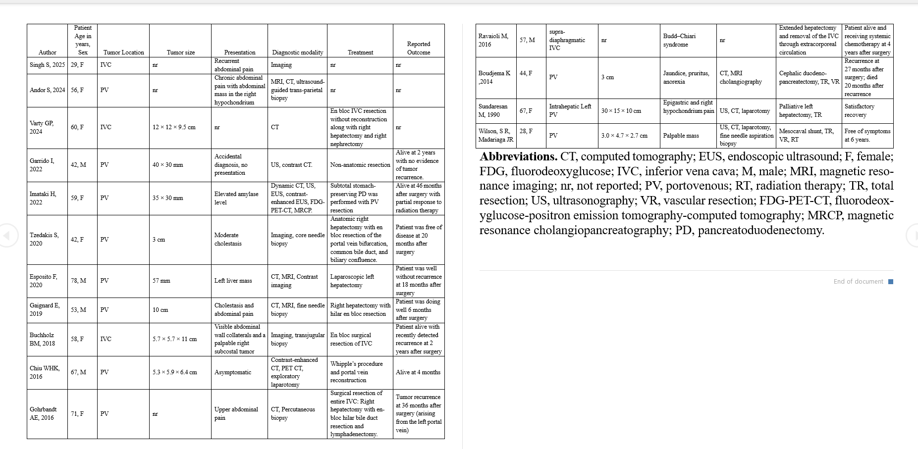
Mohammed Abusuliman, MD1, Abdullah Olimy, 2, Jonathan A. Montrose, DO3, Omar Abbas, MD3, Remy Arwani, MD1, Amr Abusuliman, MD4, Agustin Gavidia Rosario, MD3, Sumit Singla, MD3, Tobias Zuchelli, 1, Mazen Elatrache, MD3 1Henry Ford Hospital, Detroit, MI; 2Menoufia University, El-Bajour, Al Minufiyah, Egypt; 3Henry Ford Health, Detroit, MI; 4Tanta University, Qutour, Al Gharbiyah, Egypt Introduction: Leiomyosarcomas (LMS) are rare malignant tumors originating from smooth muscle tissue, most frequently occurring in the uterus, retroperitoneum, and gastrointestinal tract. They account for approximately 10%–20% of all soft tissue sarcomas and are most diagnosed in women in their sixth decade of life. LMS involving the portal vein is exceptionally rare. In this report, we present a rare case of portocaval LMS in a 38-year-old man—the youngest male patient reported to date. The diagnosis was established using endoscopic ultrasound-guided fine-needle biopsy (EUS-FNB), marking the first documented use of EUS in diagnosing such a rare intra-abdominal tumor. Table 1 summarizes prior documented case of leiomyosarcoma of the portocaval system.
Case Description/
Methods: A 38-year-old man presented to the ED with severe right upper quadrant abdominal pain radiating to the chest and ribs that started 2 weeks prior. Additional symptoms included nausea, intermittent rectal bleeding, and a 20-pound weight loss. Initial labs were unremarkable. Contrast-enhanced computed tomography (CT) showed a 4.9 × 4.0 cm heterogeneous mass abutting the second portion of the duodenum. Magnetic resonance imaging (MRI) and repeat CT revealed interval growth of a portocaval mass displacing adjacent structures and compressing the intrahepatic IVC and renal veins displacing the head of the pancreas and the second portion of the duodenum.
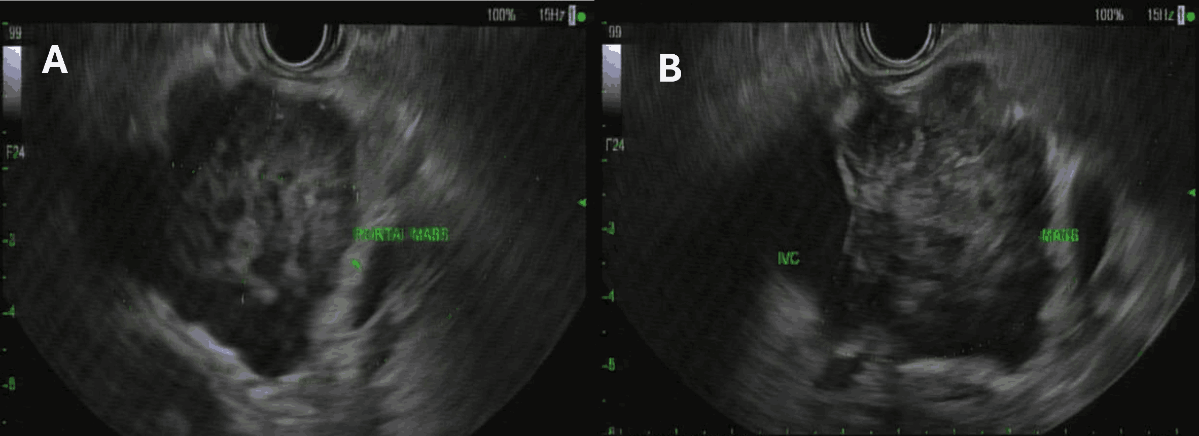
Tumor markers were within normal limits. Positron emission tomography (PET)-CT scan demonstrated mildly increased fluorodeoxyglucose uptake in the lesion. EUS identified a 4 cm hypoechoic, ill-defined mass between the duodenum, liver, and pancreas, with sonographic evidence of invasion into the portal vein and IVC. The lesion appeared distinct from the liver, pancreas, and duodenum (Figures 1A & 1B). EUS-FNB was performed, and histopathology revealed pleomorphic spindle cells with hyperchromatic nuclei, atypical mitoses, and focal necrosis. Immunohistochemistry was positive for desmin, smooth muscle actin (SMA), MIB-1 (~70%), and CD34—confirming LMS. The patient was referred for multidisciplinary evaluation by surgical oncology, vascular surgery, and medical oncology. Discussion: Our case represents the first reported instance in which EUS-FNB was used to diagnose LMS in the portocaval system, highlighting its pivotal role in identifying rare intra-abdominal malignancies, offering a minimally invasive alternative to more invasive diagnostic methods such as surgery or percutaneous biopsies.
Figure: Figure 1. Endoscopic guided ultrasonographic imaging of patient with porto-venous leiomyosarcoma. Two views (A and B) of a 4 cm hypoechoic, ill-defined portocaval mass situated between the second portion of the duodenum.
Figure: Table.1 Demographic features, clinical characteristics, treatment and diagnostic approach, and outcomes reported in published cases of leiomyosarcoma of the portovenous system
Disclosures: Mohammed Abusuliman indicated no relevant financial relationships. Abdullah Olimy indicated no relevant financial relationships. Jonathan Montrose indicated no relevant financial relationships. Omar Abbas indicated no relevant financial relationships. Remy Arwani indicated no relevant financial relationships. Amr Abusuliman indicated no relevant financial relationships. Agustin Gavidia Rosario indicated no relevant financial relationships. Sumit Singla: Boston Scientific. – Consultant. Tobias Zuchelli: Boston Scientific – Consultant. Mazen Elatrache indicated no relevant financial relationships.
Mohammed Abusuliman, MD1, Abdullah Olimy, 2, Jonathan A. Montrose, DO3, Omar Abbas, MD3, Remy Arwani, MD1, Amr Abusuliman, MD4, Agustin Gavidia Rosario, MD3, Sumit Singla, MD3, Tobias Zuchelli, 1, Mazen Elatrache, MD3. P3005 - Diagnostic Value of Endoscopic Ultrasound in a Rare Case of Portocaval Leiomyosarcoma, ACG 2025 Annual Scientific Meeting Abstracts. Phoenix, AZ: American College of Gastroenterology.