Kelly Christina. Fuller, BS1, Amanda Renee. Eukovich, DO2, Gautam Anand, MD1, Aryama Sharma, MD3 1Broward Health Medical Center, Fort Lauderdale, FL; 2Broward Health Medical Center, Pompano Beach, FL; 3Broward Health North, Deerfield Beach, FL Introduction: A Mallory-Weiss tear (MWT) is a laceration that occurs in the inner lining of the esophagus due to increased intra-abdominal pressure, most commonly due to forceful vomiting. These lesions typically occur at the gastroesophageal (GE) junction. However, tears located in the middle esophagus are exceedingly rare and underreported. Such atypical presentations pose unique diagnostic and therapeutic challenges, often associated with an increased risk of significant bleeding if diagnosis is delayed.
Case Description/
Methods: A 48-year-old man with a history of GI bleeding secondary to esophageal ulcers in the setting of Los Angeles Classification Grade D (LACD) esophagitis presented with episodes of coffee-ground emesis, dysphagia, retrosternal chest pain, and epigastric discomfort. Initial EGD revealed LACD esophagitis without evidence of ulceration or active bleeding. Cardiac workup was unremarkable, and hemoglobin remained stable. The patient was subsequently discharged.
Two days later, he was readmitted with severe retrosternal chest pain following abrupt vomiting, now complicated by hematemesis. Due to a hemoglobin of 6.7 g/dL and hemodynamic instability, he was transferred to the ICU and received an emergent transfusion. EGD demonstrated a large, five-centimeter-deep esophageal tear extending from the mid-to-lower esophagus, which was successfully managed with five endoscopic clips.
The patient remained intubated in the ICU, and hemoglobin levels stabilized. Cardiothoracic surgery was consulted; however, a full-thickness perforation was not suspected, given the patient’s clinical stability. A follow-up gastrografin esophagram showed no evidence of perforation. The patient was discharged in stable condition with a repeat EGD scheduled in 8 weeks. Discussion: This case highlights an uncommon presentation of a Mallory-Weiss tear located in the mid-esophagus, an unusual location for such lesions. While Mallory-Weiss tears at the GE junction are often self-limited, atypical locations may delay diagnosis if symptoms are not yet significant. In our case, timely EGD enabled the successful identification and treatment of the mid-esophageal tear. The patient was stabilized, avoiding progression to full-thickness esophageal rupture. Clinicians should consider atypical MWT locations in the differential diagnosis of upper GI bleeding, particularly when initial imaging or endoscopy does not reveal a lesion in the usual location. The clinical significance of mid-esophageal tears requires further investigation.
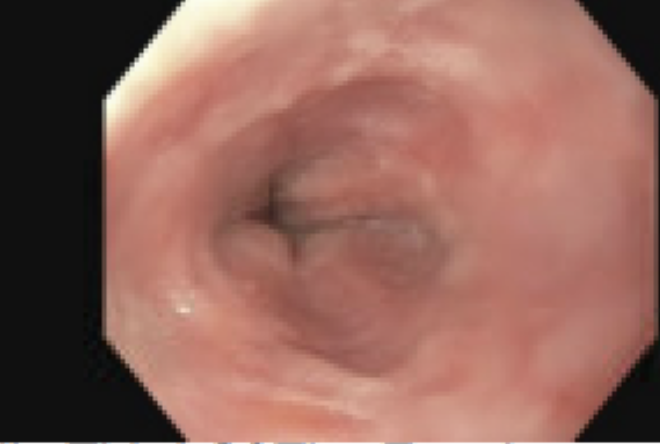
Figure: Figure 1. Initial EGD with LACD esophagitis seen in the middle-third esophagus.
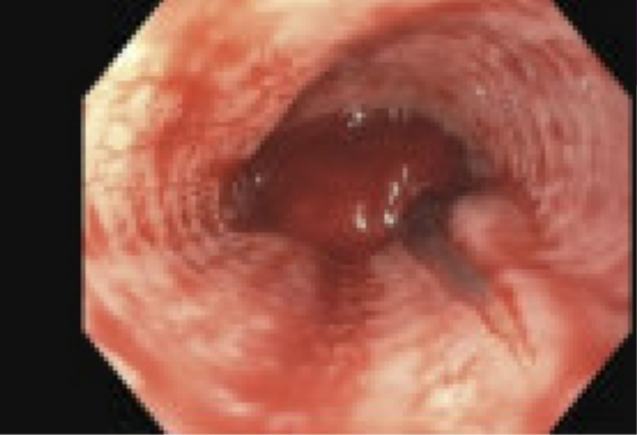
Figure: Figure 2. EGD two days later with a 5 cm Mallory-Weiss tear with adherent blood clot seen in the middle-third esophagus.
Disclosures: Kelly Fuller indicated no relevant financial relationships. Amanda Eukovich indicated no relevant financial relationships. Gautam Anand indicated no relevant financial relationships. Aryama Sharma indicated no relevant financial relationships.
Kelly Christina. Fuller, BS1, Amanda Renee. Eukovich, DO2, Gautam Anand, MD1, Aryama Sharma, MD3. P2866 - Life-Threatening Mid-Esophageal Mallory-Weiss Tear: An Uncommon Location for a Common Lesion, ACG 2025 Annual Scientific Meeting Abstracts. Phoenix, AZ: American College of Gastroenterology.