Corewell Health William Beaumont University Hospital Royal Oak, MI
Ahmed Aref, MD1, Judy Sheffeh, MD2, Mohammad Ali Sheffeh, MD3, Mujtaba Moazzam, MD1, Nishant Aggarwal, MD1, Fady Banno, MD1, Mohammed Alkhero, MD4, Ketan Rana, MD1 1Corewell Health William Beaumont University Hospital, Royal Oak, MI; 2Henry Ford Warren, Sterling Heights, MI; 3Henry Ford Warren, Warren, MI; 4Corewell Health William Beaumont University Hospital, Sterling Heights, MI Introduction: TEE is commonly used to guide structural cardiac interventions but carries a risk of esophageal injury. Despite its extensive use, there is scarce data concerning its safety. TEE can result in serious complications which can sometimes be fatal. Upper gastrointestinal injuries are the most described complications of TEE.
Case Description/
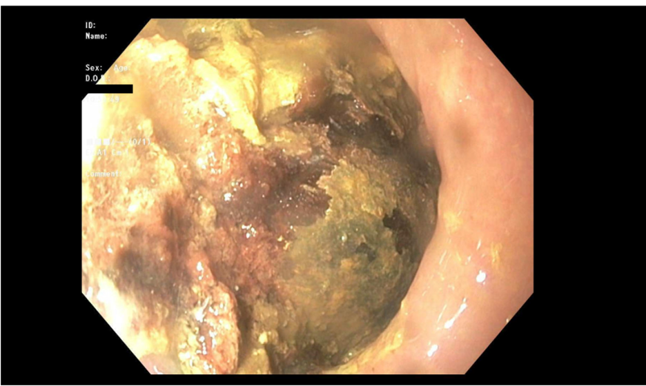
Methods: A 79-year-old female with a history of severe mitral regurgitation underwent Transcatheter Edge-to-Edge Repair (TEER) of the mitral valve. She developed postoperative atrial fibrillation that required apixaban initiation. Two days later, she presented with dysphagia, chest discomfort, and regurgitation. Esophagogastroduodenoscopy (EGD) revealed a 5 cm tear in the middle esophagus with an overlying hematoma obstructing the lumen and two diverticula near the hematoma (figure 1). Afterwards, she was able to advance her diet, and apixaban was resumed.
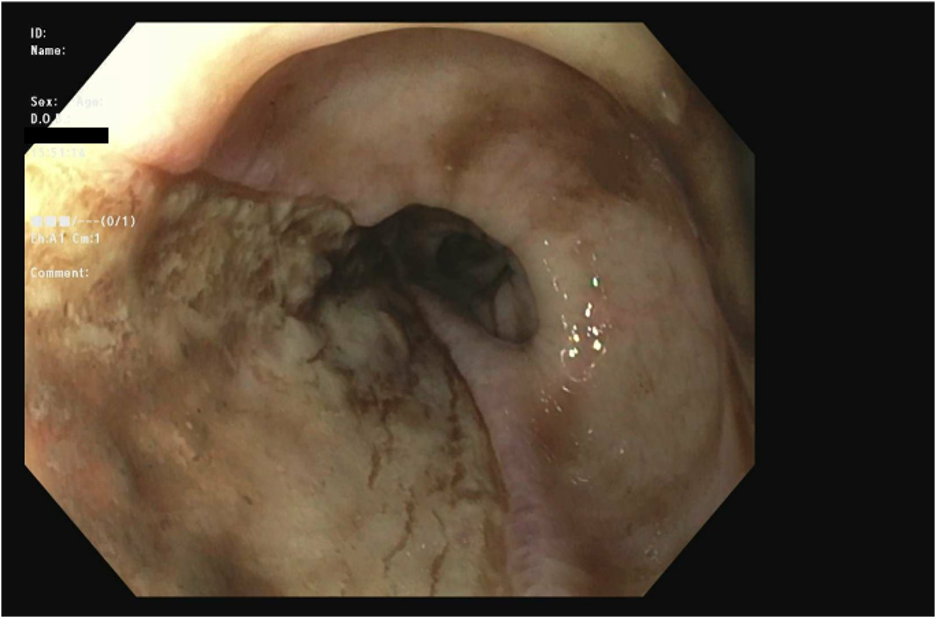
One week later, she returned with hematemesis and melena. Hemoglobin dropped from 13 g/dL to 9 g/dL. Computed tomography (CT) angiography of the thorax did not show active bleeding. A second EGD revealed a clean-based esophageal ulcer (Forrest III), from ruptured hematoma (Figure 2). Persistent hematemesis and melena prompted a third EGD, which revealed the previously seen ulcer oozing blood (Forrest IB). No definitive bleeding source was identified, and a fully covered self-expanding metal stent was placed, achieving hemostasis. Her course was further complicated by acute cholecystitis, managed with cholecystostomy due to medical instability. Despite these measures, she continued to experience bleeding, necessitating transfusions. Repeat CT angiography of the thorax showed no active bleeding or evidence of an aorto-enteric fistula. She subsequently developed septic shock requiring vasopressors. Despite aggressive management, she deteriorated. After goals-of-care discussions, she was compassionately extubated and passed away. Discussion: TEE associated esophageal injuries are underreported. Risk factors include esophageal abnormalities, anticoagulation, advanced age, and procedural complexity. Symptoms include chest pain, dysphagia, and gastrointestinal bleeding. CT is the first imaging modality to diagnose. Endoscopy with or without ultrasound and MRI, are other options for further evaluation. Esophageal hematomas are typically managed conservatively through withholding oral intake, acid suppressants, serial imaging and reversing coagulopathy.
Figure: Firs EGD showing a tear in the middle esophagus with an overlying hematoma obstructing the lumen
Figure: Second EGD revealing a clean-based esophageal ulcer (Forrest III) from ruptured hematoma
Disclosures: Ahmed Aref indicated no relevant financial relationships. Judy Sheffeh indicated no relevant financial relationships. Mohammad Ali Sheffeh indicated no relevant financial relationships. Mujtaba Moazzam indicated no relevant financial relationships. Nishant Aggarwal indicated no relevant financial relationships. Fady Banno indicated no relevant financial relationships. Mohammed Alkhero indicated no relevant financial relationships. Ketan Rana indicated no relevant financial relationships.
Ahmed Aref, MD1, Judy Sheffeh, MD2, Mohammad Ali Sheffeh, MD3, Mujtaba Moazzam, MD1, Nishant Aggarwal, MD1, Fady Banno, MD1, Mohammed Alkhero, MD4, Ketan Rana, MD1. P2860 - Esophageal Hematoma with Spontaneous Ulceration: A Rare Complication After Transesophageal Echocardiography (TEE), ACG 2025 Annual Scientific Meeting Abstracts. Phoenix, AZ: American College of Gastroenterology.