Aidan J. David, BS1, Sophie Nguyen, BS2, Joseph David, MD3, Brian M.. Fung, MD4 1Case Western Reserve University, Cleveland, OH; 2University of Arizona College of Medicine, Phoenix, Phoenix, AZ; 3University of Arizona College of Medicine, Phoenix, AZ; 4Arizona Digestive Health, Mesa, AZ Introduction: Esophagitis dissecans superficialis (EDS), also known as sloughing esophagitis, is an uncommon benign endoscopic finding characterized by desquamation of the superficial esophageal mucosa. Histology typically shows peeling sheets of sloughed mucosa over preserved basal epithelium. Patients are usually asymptomatic or present with dysphagia. Here, we present a case of EDS with an unusual presenting symptom of recurrent bleeding and a distinct endoscopic appearance lacking classic mucosal sloughing.
Case Description/
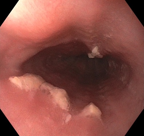
Methods: A 38-year-old man with a history of poorly controlled type 2 diabetes mellitus (A1c 8.0%) and obesity (BMI 42) presented to the gastroenterology clinic with persistent burning chest pain and frequent episodes of waking up with dried blood in his mouth. He denied dysphagia, odynophagia, or dermatologic or autoimmune disorders aside from diabetes. Esophagogastroduodenoscopy (EGD) was performed and revealed raised white lesions localized to the mid-esophagus at approximately 30 cm from the incisors (Figure). Notably, there were no strips of peeling mucosa or denudation typically associated with EDS. Biopsies of the esophageal lesions demonstrated superficial necrotic squamous epithelium detached from an otherwise unremarkable basal layer, consistent with EDS. The patient was started on omeprazole 20 mg daily and noticed improvement in reflux symptoms. Discussion: This case illustrates an atypical presentation of EDS, notable both for the symptom of recurrent oral bleeding and the deviation from its classic endoscopic appearance. Discrete raised white lesions were noted rather than desquamation or sloughing mucosa. While the etiology of EDS remains poorly defined, associations with autoimmune conditions, medications, and systemic diseases such as diabetes have been reported. Although our patient had no documented dermatologic disease, his diabetes may represent an underlying immune-mediated predisposition. Given the benign and self-limited nature of EDS, treatment is generally supportive and aimed at symptom control. This case expands the clinical and endoscopic spectrum of EDS and underscores the importance of considering it in patients with atypical esophageal findings and unexplained upper gastrointestinal bleeding.
Figure: Raised white lesions localized to the mid-esophagus
Disclosures: Aidan David indicated no relevant financial relationships. Sophie Nguyen indicated no relevant financial relationships. Joseph David indicated no relevant financial relationships. Brian Fung: Ipsen – Advisory Committee/Board Member.
Aidan J. David, BS1, Sophie Nguyen, BS2, Joseph David, MD3, Brian M.. Fung, MD4. P2842 - An Unusual Case of Esophagitis Dissecans Superficialis, ACG 2025 Annual Scientific Meeting Abstracts. Phoenix, AZ: American College of Gastroenterology.