
Monday Poster Session
Category: Colon

Muhammad Sohaib, MD
UCHealth Parkview Medical Center
Pueblo, CO
A 51-year-old woman with no prior GI screenings was admitted for unrelated orthopedic issues and found to have profound anemia (Hb 5.1 g/dL). She denied overt GI bleeding but reported months of fatigue and dizziness. Iron studies showed ferritin 2 ng/mL and iron saturation 4%. After receiving 3 units of pRBC, she was discharged on iron supplements and referred to GI to be followed up in an outpatient setting.
Initial EGD revealed only a hiatal hernia; colonoscopy was repeated due to poor preparation. On the second attempt, four 3–4 mm sessile polyps were removed from the sigmoid colon using a cold snare. Diverticulosis and grade 1 internal hemorrhoids were also noted. Biopsies from the sigmoid colon revealed one tubular adenoma and a pyogenic granuloma. The patient was subsequently discharged from the hospital. Hemoglobin remained stable at the 3-month follow-up without further bleeding.
Discussion:
PG, also called lobular capillary hemangioma, is a rapidly growing lesion that may bleed with minimal trauma. Though common on the skin, PG is rarely reported in the GI tract, where it can mimic other vascular or neoplastic lesions. Diagnosis requires histological confirmation, as appearance may overlap with Kaposi sarcoma or angiomatosis.
While GI involvement is uncommon, several case reports highlight PG as a cause of occult or overt GI bleeding. Meyer-Herbon et al. described a recurrent sigmoid PG requiring multiple resections. Ikeoka et al. reported jejunal PG after variceal banding, while Martinez et al. noted duodenal PG with a resolution of anemia post-resection.
Our case adds to the limited literature on colonic PG and reinforces the importance of considering vascular lesions in unexplained iron deficiency anemia. With increasing endoscopic utilization, PG may be under-recognized rather than truly rare. Greater awareness and reporting are needed to define its prevalence and guide optimal management.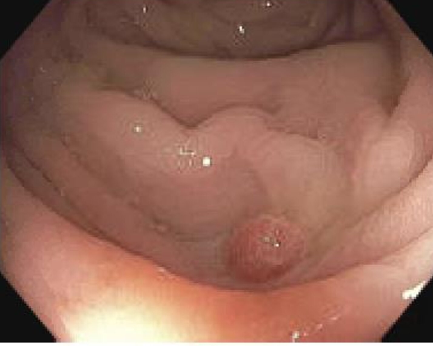
Figure: Endoscopic view of a polypoid lesion in the sigmoid colon later identified as pyogenic granuloma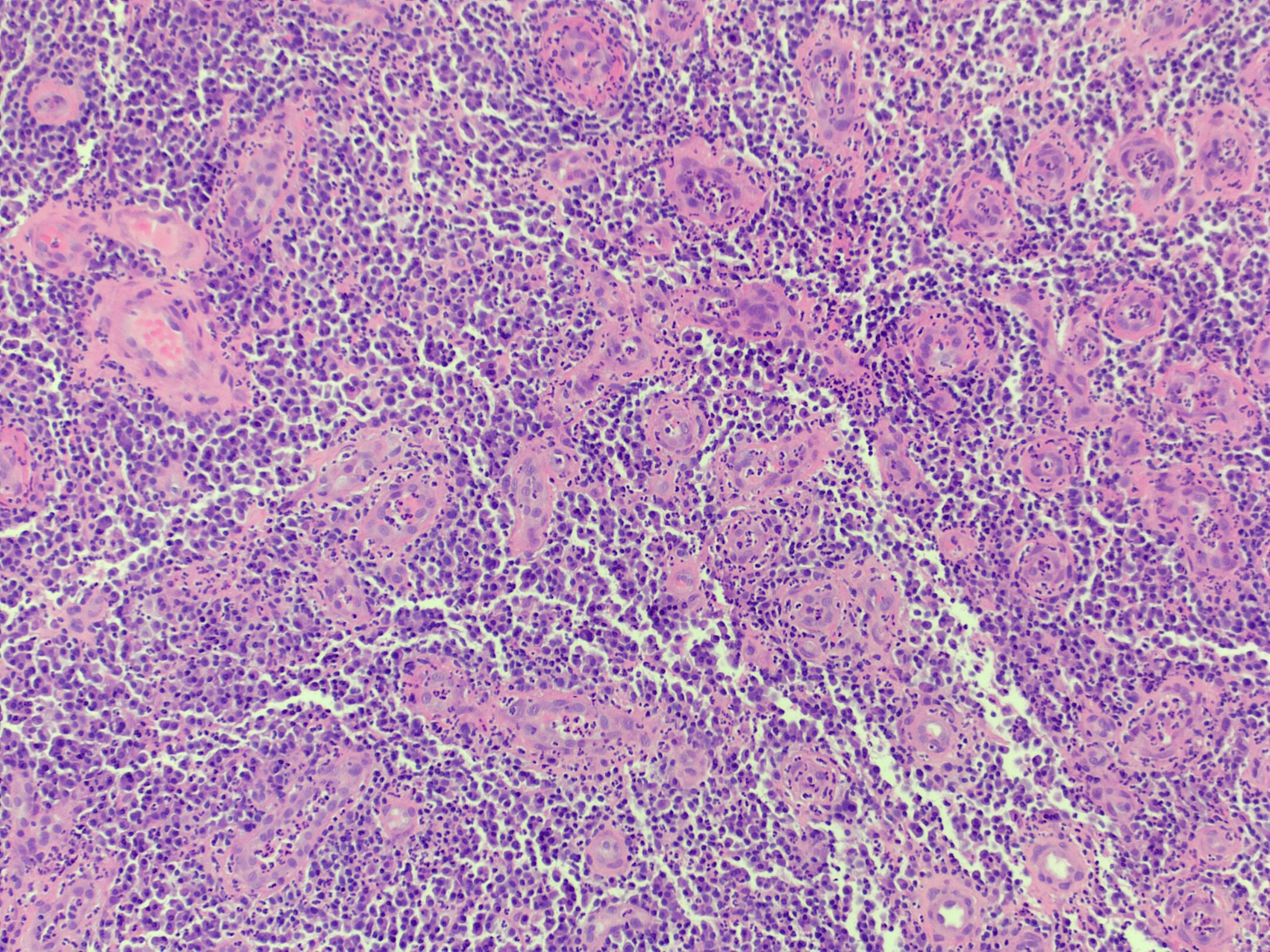
Figure: Histologic section showing lobular proliferation of capillaries consistent with pyogenic granuloma
Disclosures:
Muhammad Sohaib indicated no relevant financial relationships.
Mustafa Nayeem indicated no relevant financial relationships.
Rajiv N. Singh indicated no relevant financial relationships.
Muhammad Shammas Tariq indicated no relevant financial relationships.
Fatima Zia indicated no relevant financial relationships.
Zainab Zia indicated no relevant financial relationships.
Sara Ahmed indicated no relevant financial relationships.
Muhammad Hamza indicated no relevant financial relationships.
Muhammad Sohaib, MD1, Mustafa Nayeem, MD2, Rajiv N. Singh, MBBS2, Muhammad Shammas Tariq, MBBS3, Fatima Zia, MD4, Zainab Zia, MD5, Sara Ahmed, MD6, Muhammad Hamza, MD7. P2524 - Not Just a Polyp: Unmasking a Rare Bleeding Lesion Treated Endoscopically, ACG 2025 Annual Scientific Meeting Abstracts. Phoenix, AZ: American College of Gastroenterology.