Priscilla Lajara, MD1, Joseph Marte, MD1, Mohamed Farag, MD2, Dogmin Shin, MD1, Elona Shehi, MD1 1BronxCare Health System, Bronx, NY; 2Bronxcare, Bronx, NY Introduction: Filiform Polyposis (FP), also known as pseudo polyposis, is a benign, non-neoplastic, rare type of inflammatory polyposis associated with inflammatory bowel disease (IBD). We present a rare case of a 65-year-old male with no IBD with multiple filiform polyps.
Case Description/
Methods: A 65-year-old male with a medical history of HLD, presented to the GI clinic for a surveillance colonoscopy. On evaluation, he denied abdominal pain, constipation, diarrhea, hematemesis, or hematochezia. He also denied weight loss, anorexia, family history of GI malignancies, or chronic GI disorders. Ten years earlier, he had his first screening colonoscopy that revealed multiple finger-like projections in the descending and transverse colon. Otherwise, the colonic mucosa appeared normal. He had three more colonoscopies, showing an increase in the size, number, and extent of the polyps. In the latest colonoscopy, almost the entire colon was affected, sparing only the caecum and distal rectum. Largest polyps were removed with cold and hot snare. The pathology showed benign colonic projections of fibrovascular submucosa containing ganglion cells consistent with FP. The remaining of the colonic mucosa was normal.
Discussion: Filiform Polyposis is a rare inflammatory polyposis most commonly associated with IBD. It is a benign worm slender-like submucosal projection composed of a fibrovascular stalk found in normal colonic tissue without evidence of dysplasia. It is strongly associated with ulcerative colitis and Crohn's disease. It can be found in the small and large intestines, with the vast majority in the sigmoid colon. The prevalence of FP in non-IBD is not known, and there are a scarce number of cases of FP in patients with no history of inflammatory bowel disease. FP has been associated with non-IBD conditions such as necrotizing enterocolitis, enema-induced colitis, stercoral ulcer, Langerhans cell histiocytosis X, and colonic tuberculosis.
The exact pathogenesis of FP is not clearly understood. Most cases are associated with post-inflammatory periods of mucosal repair where growth factors form residual pedunculated stocks of normal mucosa. FP sizes and shapes vary and can be difficult to distinguish from other inflammatory processes.
FP is not considered precancerous, however, cases of adenocarcinoma have been reported. FP can present with similar symptoms as IBD, and, if polyps conglomerate, can cause obstructive mass and intussusception. FB in non-IBD patients has been shown to have a benign clinical course.
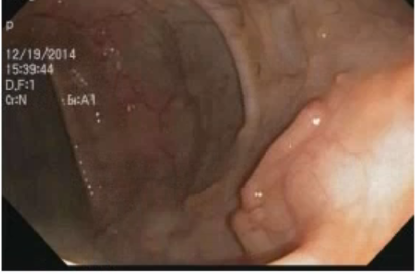
Figure: First screening colonoscopy showing finger-like polyps in the transverse colon
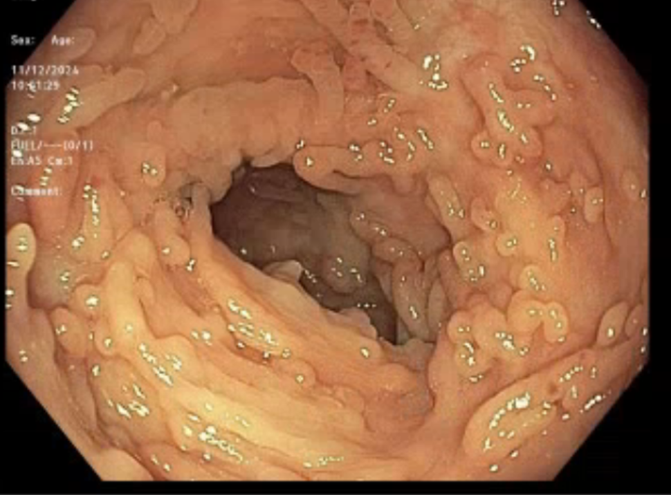
Figure: Colonoscopy 2024, Florid Polyposis in the descending colon
Disclosures: Priscilla Lajara indicated no relevant financial relationships. Joseph Marte indicated no relevant financial relationships. Mohamed Farag indicated no relevant financial relationships. Dogmin Shin indicated no relevant financial relationships. Elona Shehi indicated no relevant financial relationships.
Priscilla Lajara, MD1, Joseph Marte, MD1, Mohamed Farag, MD2, Dogmin Shin, MD1, Elona Shehi, MD1. P2511 - A Case of Filiform Polyposis in a Non-IBD Patient, ACG 2025 Annual Scientific Meeting Abstracts. Phoenix, AZ: American College of Gastroenterology.

 photo")