University of Illinois College of Medicine Chicago, IL
Yousif Esho, MD1, Hetal Patel, MD1, Dexter E. Nwachukwu, DO1, James S. Love, MD1, Wadih Chacra, MD1, Adam E. Mikolajczyk, MD2 1University of Illinois College of Medicine, Chicago, IL; 2University of Illinois, Chicago, IL Introduction: Biliary tract cancers are uncommon, with the vast majority being adenocarcinomas. Squamous cell carcinoma (SCC) of the gallbladder or bile ducts is exceedingly rare, accounting for less than 1% of all biliary tumors. This histologic subtype is poorly understood and associated with a more aggressive clinical course and higher mortality. Due to its rarity, there are no established treatment guidelines, and it is often managed similarly to adenocarcinoma. There are many risk factors and conditions associated with biliary malignancies; however, many cases have no clear etiology.
Case Description/
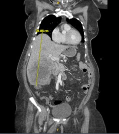
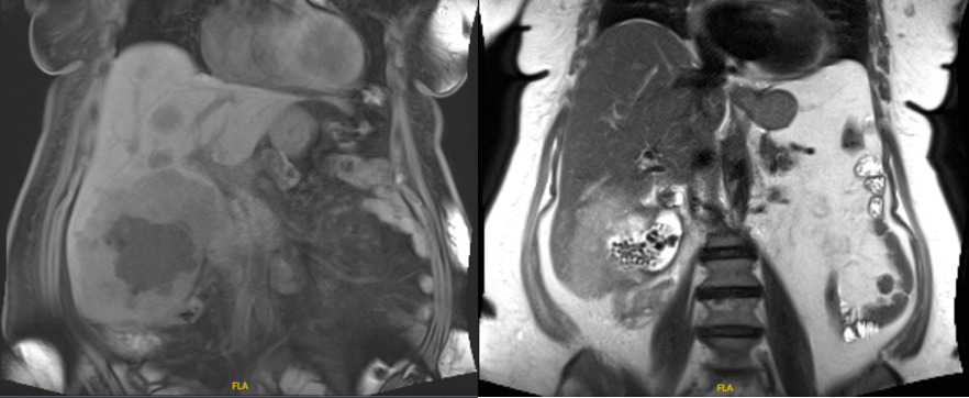
Methods: We present a case of a 76-year-old woman with no history of chronic liver disease who was initially evaluated by her primary care physician for lip swelling. She was referred to the emergency department for suspected angioedema, where a physical examination incidentally revealed hepatomegaly. CT imaging showed multiple hypodense hepatic lesions (Figure 1) with MRI revealing the largest measuring 8.0 x 7.6 x 7.3 cm in the right hepatic lobe, contiguous with the gallbladder lumen, which contained multiple stones (Figure 2). Biopsy of the dominant hepatic mass confirmed metastatic squamous cell carcinoma, with the gallbladder presumed to be the primary site as cross-sectional imaging was negative for alternative sources of primary malignancy.
Within one month of diagnosis, the patient completed the first day of her chemotherapy regimen including folinic acid, fluorouracil, and oxaliplatin. Unfortunately, her course was complicated by septic shock, metabolic acidosis requiring hemodialysis, and disseminated intravascular coagulation. Despite intensive care, the patient died from cardiac arrest due to pulseless electrical activity. Discussion: This case demonstrates the aggressiveness of squamous cell carcinoma of the gallbladder, known for its rapid progression, extensive local invasion, and poor outcomes. The etiology is not understood, but chronic inflammation of the biliary tree is hypothesized to cause squamous metaplasia and subsequent dysplasia. Clinically and radiographically, it is indistinguishable from adenocarcinomas, with histologic analysis as the only way to confirm the diagnosis. Management is modeled after adenocarcinoma, with surgical resection as the mainstay, and chemotherapy or radiation considered as adjuncts. This case adds to the limited literature on biliary SCC and highlights the importance of reporting such rare variants to inform future diagnostic and therapeutic approaches.
Figure: Figure 1 - CT imaging showing multiple hypodense lesions in the liver and subsequent hepatomegaly.
Figure: Figure 2 - MRI showing evidence of the largest hepatic lesion being contiguous with the gallbladder lumen which contained multiple stones.
Disclosures: Yousif Esho indicated no relevant financial relationships. Hetal Patel indicated no relevant financial relationships. Dexter Nwachukwu indicated no relevant financial relationships. James Love indicated no relevant financial relationships. Wadih Chacra indicated no relevant financial relationships. Adam Mikolajczyk indicated no relevant financial relationships.
Yousif Esho, MD1, Hetal Patel, MD1, Dexter E. Nwachukwu, DO1, James S. Love, MD1, Wadih Chacra, MD1, Adam E. Mikolajczyk, MD2. P2327 - Squamous Cell Carcinoma of the Gallbladder: A Rare and Aggressive Entity, ACG 2025 Annual Scientific Meeting Abstracts. Phoenix, AZ: American College of Gastroenterology.