P4525 - A Patient With Pancreatic Cancer Following Radiation Therapy for Mucosa-Associated Lymphoid Tissue (MALT) Lymphoma Diagnosed Incidentally by Upper Endoscopy
Mikaela Lipp, BS1, Robert Klein, MD, FACG2, Michael Shapiro, MD3, Keith Sultan, MD, FACG2 1Duke University, Durham, NC; 2Zucker School of Medicine Hofstra University, Hempstead, NY; 3Northwell Health, New Hyde Park, NY Introduction: Pancreatic cancer and gastric mucosa-associated lymphoid tissue (MALT) lymphoma are unrelated malignancies. However, there are reports of pancreatic cancer developing following radiation therapy (RT) for MALT lymphoma, as the pancreas is within the radiation field.1 We present a case of pancreatic cancer, incidentally diagnosed by upper endoscopy (EGD), occurring years after radiation therapy for MALT lymphoma.
Case Description/
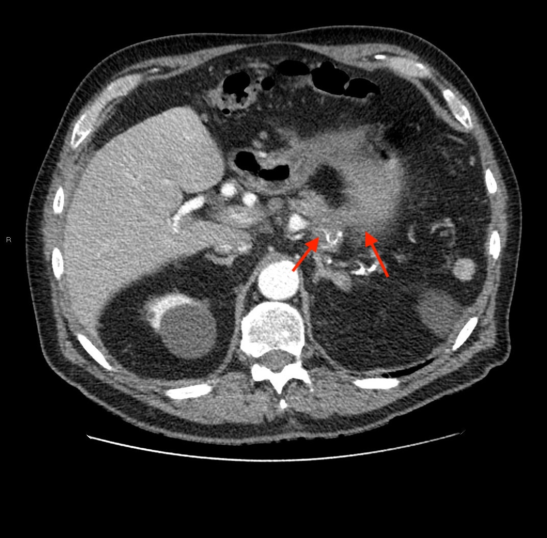
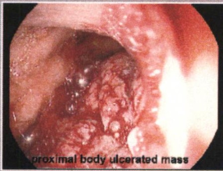
Methods: A 72-year-old asymptomatic man presented for a surveillance EGD for a history of both intestinal metaplasia in the stomach and non-H. pylori-associated gastric MALT lymphoma treated with RT 17 years prior. The EGD was significant for an ulcerated, hemorrhagic mass in the proximal gastric body (Figure 1). Given the patient’s history, this was presumed to be recurrent MALT lymphoma. However, biopsies revealed invasive poorly differentiated adenocarcinoma with micropapillary features consistent with pancreaticobiliary origin. The patient underwent CT scan of the abdomen which revealed a 4.3 x 3.7 cm heterogenous mass within the pancreatic body/tail extending along the the lesser curvature of the stomach. Numerous soft tissue nodular densities were noted along the anterior omentum/mesentery compatible with carcinomatosis (Figure 2). Ca 19-9 was markedly elevated at 3708. The patient was referred to oncology and treated with systemic chemotherapy. Discussion: The patient described above has two interesting facets to his presentation. Firstly, he developed pancreatic cancer 17 years after receiving RT for MALT lymphoma. This is a rare complication of this treatment.1 Secondly, the cancer was diagnosed as a gastric mass noted incidentally on EGD. There are rare case reports of the diagnosis being made in this fashion.2 Our experience reinforces the importance of a thorough patient history and awareness of the risks of previous RT. Additionally, pancreatic cancer should be included in the differential diagnosis of endoscopically noted gastric masses.
References:
1. Nam, H et al. Long-Term Clinical Outcome and Predictive Factors for Relapse after Radiation Therapy in 145 Patients with Stage I Gastric B-Cell Lymphoma of Mucosa-Associated Lymphoid Tissue Type. Cancers. 2021; vol. 13,2 169.
2. Chelimilla H et al. Rare endoscopic manifestation of pancreatic adenocarcinoma. Case Rep Gastroenterol. 2012;6(2):496-501
Figure: Figure 1 - Endoscopic view of an ulcerated, hemorrhagic mass seen in the proximal gastric body
Figure: Figure 2 - CT image of a pancreatic mass extending across the lesser curvature of the stomach
Disclosures: Mikaela Lipp indicated no relevant financial relationships. Robert Klein indicated no relevant financial relationships. Michael Shapiro indicated no relevant financial relationships. Keith Sultan indicated no relevant financial relationships.
Mikaela Lipp, BS1, Robert Klein, MD, FACG2, Michael Shapiro, MD3, Keith Sultan, MD, FACG2. P4525 - A Patient With Pancreatic Cancer Following Radiation Therapy for Mucosa-Associated Lymphoid Tissue (MALT) Lymphoma Diagnosed Incidentally by Upper Endoscopy, ACG 2025 Annual Scientific Meeting Abstracts. Phoenix, AZ: American College of Gastroenterology.